 Movie
Movie Controller

[English] 日本語
 Yorodumi
- PDB-4l1g: Crystal structure of the Bc1960 peptidoglycan N-acetylglucosamine... -
+ Open data

- Basic information
| Entry | Database: PDB / ID: 4l1g | ||||||
|---|---|---|---|---|---|---|---|
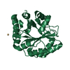
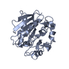
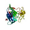
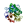
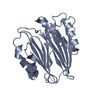
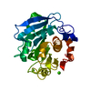
| Title | Crystal structure of the Bc1960 peptidoglycan N-acetylglucosamine deacetylase from Bacillus cereus | ||||||
 Components Components | Peptidoglycan N-acetylglucosamine deacetylase | ||||||
 Keywords Keywords | HYDROLASE / tim barrel / polysaccharide deacetylase | ||||||
| Function / homology |  Function and homology information Function and homology informationpeptidoglycan-N-acetylglucosamine deacetylase / hydrolase activity, acting on carbon-nitrogen (but not peptide) bonds / carbohydrate metabolic process / metal ion binding Similarity search - Function | ||||||
| Biological species |  | ||||||
| Method |  X-RAY DIFFRACTION / SYNCHROTRON / MOLECULAR REPLACEMENT / Resolution: 2.336 Å X-RAY DIFFRACTION / SYNCHROTRON / MOLECULAR REPLACEMENT / Resolution: 2.336 Å | ||||||
 Authors Authors | Tsalafouta, A. / Fadouloglou, V.E. / Kokkinidis, M. | ||||||
 Citation Citation | Journal: J.Am.Chem.Soc. / Year: 2017 Title: Unusual alpha-Carbon Hydroxylation of Proline Promotes Active-Site Maturation. Authors: Fadouloglou, V.E. / Balomenou, S. / Aivaliotis, M. / Kotsifaki, D. / Arnaouteli, S. / Tomatsidou, A. / Efstathiou, G. / Kountourakis, N. / Miliara, S. / Griniezaki, M. / Tsalafouta, A. / ...Authors: Fadouloglou, V.E. / Balomenou, S. / Aivaliotis, M. / Kotsifaki, D. / Arnaouteli, S. / Tomatsidou, A. / Efstathiou, G. / Kountourakis, N. / Miliara, S. / Griniezaki, M. / Tsalafouta, A. / Pergantis, S.A. / Boneca, I.G. / Glykos, N.M. / Bouriotis, V. / Kokkinidis, M. | ||||||
| History |
|
- Structure visualization
| Structure viewer | Molecule: MolmilJmol/JSmol |
|---|
- Downloads & links
-Download
| PDBx/mmCIF format | 4l1g.cif.gz | 362.7 KB | Display | PDBx/mmCIF format |
|---|---|---|---|---|
| PDB format | pdb4l1g.ent.gz | 296.4 KB | Display | PDB format |
| PDBx/mmJSON format | 4l1g.json.gz | Tree view | PDBx/mmJSON format | |
| Others |  Other downloads Other downloads |
-Validation report
| Arichive directory | https://data.pdbj.org/pub/pdb/validation_reports/l1/4l1gftp://data.pdbj.org/pub/pdb/validation_reports/l1/4l1g | HTTPS FTP |
|---|
-Related structure data
| Related structure data |  1w1bS S: Starting model for refinement |
|---|---|
| Similar structure data |


-Links
 PDBj
PDBj
- Assembly
| Deposited unit | 
| ||||||||
|---|---|---|---|---|---|---|---|---|---|
| 1 | 
| ||||||||
| 2 | 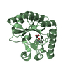
| ||||||||
| 3 | 
| ||||||||
| 4 | 
| ||||||||
| Unit cell |
|
-Components
| #1: Protein | Mass: 31448.307 Da / Num. of mol.: 4 Source method: isolated from a genetically manipulated source Source: (gene. exp.) References: UniProt: Q81EK9, Hydrolases; Acting on carbon-nitrogen bonds, other than peptide bonds; In linear amides #2: Chemical | ChemComp-SO4 /   Mass: 96.063 Da / Num. of mol.: 6 / Source method: obtained synthetically / Formula: SO4 Mass: 96.063 Da / Num. of mol.: 6 / Source method: obtained synthetically / Formula: SO4#3: Chemical | ChemComp-ACT /   Mass: 59.044 Da / Num. of mol.: 4 / Source method: obtained synthetically / Formula: C2H3O2 Mass: 59.044 Da / Num. of mol.: 4 / Source method: obtained synthetically / Formula: C2H3O2#4: Water | ChemComp-HOH / |  Mass: 18.015 Da / Num. of mol.: 769 / Source method: isolated from a natural source / Formula: H2O Mass: 18.015 Da / Num. of mol.: 769 / Source method: isolated from a natural source / Formula: H2O |
|---|
-Experimental details
-Experiment
| Experiment | Method: X-RAY DIFFRACTION / Number of used crystals: 1 |
|---|
- Sample preparation
| Crystal | Density Matthews: 2 Å3/Da / Density % sol: 38.61 % |
|---|---|
| Crystal grow | Temperature: 291 K / Method: vapor diffusion, hanging drop / pH: 4.5 Details: 15% PEG 4000, 0.1M ammonium sulfate, 0.1M sodium acetate, pH 4.5, VAPOR DIFFUSION, HANGING DROP, temperature 291K |
-Data collection
| Diffraction | Mean temperature: 100 K |
|---|---|
| Diffraction source | Source: SYNCHROTRON / Site: EMBL/DESY, HAMBURG  / Beamline: BW7A / Wavelength: 0.817 Å / Beamline: BW7A / Wavelength: 0.817 Å |
| Detector | Type: MAR CCD 165 mm / Detector: CCD / Date: Dec 15, 2009 / Details: monochromators |
| Radiation | Monochromator: Si [111] / Protocol: SINGLE WAVELENGTH / Monochromatic (M) / Laue (L): M / Scattering type: x-ray |
| Radiation wavelength | Wavelength: 0.817 Å / Relative weight: 1 |
| Reflection | Resolution: 2.336→80 Å / Num. obs: 45861 / % possible obs: 100 % / Observed criterion σ(F): 0.5 / Observed criterion σ(I): 1 |
| Reflection shell | Resolution: 2.336→2.3868 Å / % possible all: 99.5 |
- Processing
| Software |
| |||||||||||||||||||||||||||||||||||||||||||||||||||||||||||||||||||||||||||||||||||||||||||||||||||||||||||||||||||||||
|---|---|---|---|---|---|---|---|---|---|---|---|---|---|---|---|---|---|---|---|---|---|---|---|---|---|---|---|---|---|---|---|---|---|---|---|---|---|---|---|---|---|---|---|---|---|---|---|---|---|---|---|---|---|---|---|---|---|---|---|---|---|---|---|---|---|---|---|---|---|---|---|---|---|---|---|---|---|---|---|---|---|---|---|---|---|---|---|---|---|---|---|---|---|---|---|---|---|---|---|---|---|---|---|---|---|---|---|---|---|---|---|---|---|---|---|---|---|---|---|---|
| Refinement | Method to determine structure: MOLECULAR REPLACEMENT Starting model: PDB ENTRY 1W1B Resolution: 2.336→30.639 Å / SU ML: 0.23 / σ(F): 1.33 / Phase error: 23.69 / Stereochemistry target values: ML
| |||||||||||||||||||||||||||||||||||||||||||||||||||||||||||||||||||||||||||||||||||||||||||||||||||||||||||||||||||||||
| Solvent computation | Shrinkage radii: 0.9 Å / VDW probe radii: 1.11 Å / Solvent model: FLAT BULK SOLVENT MODEL | |||||||||||||||||||||||||||||||||||||||||||||||||||||||||||||||||||||||||||||||||||||||||||||||||||||||||||||||||||||||
| Refinement step | Cycle: LAST / Resolution: 2.336→30.639 Å
| |||||||||||||||||||||||||||||||||||||||||||||||||||||||||||||||||||||||||||||||||||||||||||||||||||||||||||||||||||||||
| Refine LS restraints |
| |||||||||||||||||||||||||||||||||||||||||||||||||||||||||||||||||||||||||||||||||||||||||||||||||||||||||||||||||||||||
| LS refinement shell |
|
