 Movie
Movie Controller

[English] 日本語
 Yorodumi
- PDB-5bzt: Crystal structure of the murine cd44 hyaluronan binding domain co... -
+ Open data

- Basic information
| Entry | Database: PDB / ID: 5bzt | ||||||
|---|---|---|---|---|---|---|---|
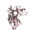
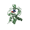
| Title | Crystal structure of the murine cd44 hyaluronan binding domain complex with a small molecule | ||||||
 Components Components | CD44 antigen | ||||||
 Keywords Keywords | PROTEIN BINDING / Link module | ||||||
| Function / homology |  Function and homology information Function and homology informationHyaluronan metabolism / Hyaluronan degradation / positive regulation of monocyte aggregation / macrophage fusion / hyaluronic acid binding / macrophage migration inhibitory factor receptor complex / Degradation of the extracellular matrix / negative regulation of regulatory T cell differentiation / Integrin cell surface interactions / regulation of lamellipodium morphogenesis ...Hyaluronan metabolism / Hyaluronan degradation / positive regulation of monocyte aggregation / macrophage fusion / hyaluronic acid binding / macrophage migration inhibitory factor receptor complex / Degradation of the extracellular matrix / negative regulation of regulatory T cell differentiation / Integrin cell surface interactions / regulation of lamellipodium morphogenesis / Cell surface interactions at the vascular wall / postsynapse organization / monocyte aggregation / wound healing involved in inflammatory response / hyaluronan catabolic process / branching involved in prostate gland morphogenesis / positive regulation of adaptive immune response / type II transforming growth factor beta receptor binding / negative regulation of mature B cell apoptotic process / negative regulation of CD4-positive, alpha-beta T cell proliferation / positive regulation of neutrophil apoptotic process / regulation of modification of postsynaptic structure / channel regulator activity / positive regulation of heterotypic cell-cell adhesion / wound healing, spreading of cells / epidermal growth factor receptor binding / cargo receptor activity / branching involved in ureteric bud morphogenesis / negative regulation of intrinsic apoptotic signaling pathway in response to DNA damage by p53 class mediator / negative regulation of DNA damage response, signal transduction by p53 class mediator / microvillus / lamellipodium membrane / cell projection / Neutrophil degranulation / cellular response to fibroblast growth factor stimulus / receptor-mediated endocytosis / T cell activation / phosphoprotein binding / regulation of cell growth / negative regulation of inflammatory response / Wnt signaling pathway / cytokine-mediated signaling pathway / neuron projection development / transmembrane signaling receptor activity / cell migration / presynapse / basolateral plasma membrane / positive regulation of ERK1 and ERK2 cascade / cell adhesion / apical plasma membrane / postsynapse / membrane raft / inflammatory response / external side of plasma membrane / positive regulation of gene expression / protein kinase binding / negative regulation of apoptotic process / glutamatergic synapse / cell surface / Golgi apparatus / protein-containing complex / extracellular region / plasma membrane / cytosol Similarity search - Function | ||||||
| Biological species |  | ||||||
| Method |  X-RAY DIFFRACTION / SYNCHROTRON / MOLECULAR REPLACEMENT / molecular replacement / Resolution: 1.25 Å X-RAY DIFFRACTION / SYNCHROTRON / MOLECULAR REPLACEMENT / molecular replacement / Resolution: 1.25 Å | ||||||
 Authors Authors | Liu, L.K. / Finzel, B.C. | ||||||
 Citation Citation | Journal: To Be Published Title: Crystal structure of the murine cd44 hyaluronan binding domain complex with a small molecule Authors: Liu, L.K. / Finzel, B.C. | ||||||
| History |
|
- Structure visualization
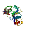
| Structure viewer | Molecule: MolmilJmol/JSmol |
|---|
- Downloads & links
-Download
| PDBx/mmCIF format | 5bzt.cif.gz | 49.4 KB | Display | PDBx/mmCIF format |
|---|---|---|---|---|
| PDB format | pdb5bzt.ent.gz | 32.6 KB | Display | PDB format |
| PDBx/mmJSON format | 5bzt.json.gz | Tree view | PDBx/mmJSON format | |
| Others |  Other downloads Other downloads |
-Validation report
| Arichive directory | https://data.pdbj.org/pub/pdb/validation_reports/bz/5bztftp://data.pdbj.org/pub/pdb/validation_reports/bz/5bzt | HTTPS FTP |
|---|
-Related structure data
| Related structure data |  5bzcC 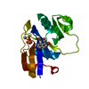 5bzeC  5bzfC 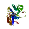 5bzgC 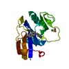 5bzhC 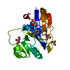 5bziC 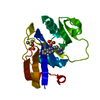 5bzjC 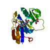 5bzkC 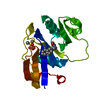 5bzlC 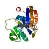 5bzmC 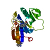 5bznC 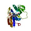 5bzoC  5bzpC 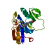 5bzqC  5bzrC 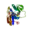 5bzsC C: citing same article ( |
|---|---|
| Similar structure data |


-Links
 PDBj
PDBj


- Assembly
| Deposited unit | 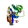
| ||||||||
|---|---|---|---|---|---|---|---|---|---|
| 1 |
| ||||||||
| Unit cell |
|
-Components
-Protein , 1 types, 1 molecules A
| #1: Protein | Mass: 16855.803 Da / Num. of mol.: 1 / Fragment: HYALURONAN BINDING DOMAIN, RESIDUES 21-171 / Mutation: H23M; Q24N Source method: isolated from a genetically manipulated source Source: (gene. exp.)  |
|---|
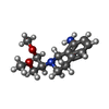
-Non-polymers , 5 types, 93 molecules 


| #2: Chemical | ChemComp-DMS /  Mass: 78.133 Da / Num. of mol.: 1 / Source method: obtained synthetically / Formula: C2H6OS / Comment: DMSO, precipitant*YM Mass: 78.133 Da / Num. of mol.: 1 / Source method: obtained synthetically / Formula: C2H6OS / Comment: DMSO, precipitant*YM | ||||
|---|---|---|---|---|---|
| #3: Chemical | ChemComp-SO4 /  Mass: 96.063 Da / Num. of mol.: 1 / Source method: obtained synthetically / Formula: SO4 Mass: 96.063 Da / Num. of mol.: 1 / Source method: obtained synthetically / Formula: SO4 | ||||
| #4: Chemical |  Mass: 250.337 Da / Num. of mol.: 2 / Source method: obtained synthetically / Formula: C14H22N2O2 Mass: 250.337 Da / Num. of mol.: 2 / Source method: obtained synthetically / Formula: C14H22N2O2#5: Chemical | ChemComp-GOL / |  Mass: 92.094 Da / Num. of mol.: 1 / Source method: obtained synthetically / Formula: C3H8O3 Mass: 92.094 Da / Num. of mol.: 1 / Source method: obtained synthetically / Formula: C3H8O3#6: Water | ChemComp-HOH / | Mass: 18.015 Da / Num. of mol.: 88 / Source method: isolated from a natural source / Formula: H2O |
-Details
| Has protein modification | Y |
|---|
-Experimental details
-Experiment
| Experiment | Method: X-RAY DIFFRACTION / Number of used crystals: 1 |
|---|
- Sample preparation
| Crystal | Density Matthews: 2.14 Å3/Da / Density % sol: 42.42 % |
|---|---|
| Crystal grow | Temperature: 298 K / Method: vapor diffusion, hanging drop / pH: 6.5 / Details: PEG MME 5000, MES, (NH4)2SO4 |
-Data collection
| Diffraction | Mean temperature: 100 K |
|---|---|
| Diffraction source | Source: SYNCHROTRON / Site: APS  / Beamline: 17-ID / Wavelength: 1 Å / Beamline: 17-ID / Wavelength: 1 Å |
| Detector | Type: DECTRIS PILATUS 6M / Detector: PIXEL / Date: Apr 18, 2015 |
| Radiation | Monochromator: Si(111) / Protocol: SINGLE WAVELENGTH / Monochromatic (M) / Laue (L): M / Scattering type: x-ray |
| Radiation wavelength | Wavelength: 1 Å / Relative weight: 1 |
| Reflection | Resolution: 1.25→5.797 Å / Num. obs: 36555 / % possible obs: 94.3 % / Redundancy: 3.4 % / Net I/σ(I): 26.6 |
| Reflection shell | Resolution: 1.25→1.254 Å / Redundancy: 3.5 % / Rmerge(I) obs: 0.102 / Mean I/σ(I) obs: 11.6 / % possible all: 91 |
-Phasing
| Phasing | Method: molecular replacement |
|---|
- Processing
| Software |
| |||||||||||||||||||||||||||||||||||||||||||||||||||||||||||||||||
|---|---|---|---|---|---|---|---|---|---|---|---|---|---|---|---|---|---|---|---|---|---|---|---|---|---|---|---|---|---|---|---|---|---|---|---|---|---|---|---|---|---|---|---|---|---|---|---|---|---|---|---|---|---|---|---|---|---|---|---|---|---|---|---|---|---|---|
| Refinement | Method to determine structure: MOLECULAR REPLACEMENT / Resolution: 1.25→5.797 Å / Cor.coef. Fo:Fc: 0.963 / Cor.coef. Fo:Fc free: 0.951 / WRfactor Rfree: 0.2014 / WRfactor Rwork: 0.191 / FOM work R set: 0.8958 / SU B: 0.617 / SU ML: 0.028 / SU R Cruickshank DPI: 0.0505 / SU Rfree: 0.0499 / Cross valid method: THROUGHOUT / σ(F): 0 / ESU R: 0.05 / ESU R Free: 0.05 / Stereochemistry target values: MAXIMUM LIKELIHOOD Details: HYDROGENS HAVE BEEN ADDED IN THE RIDING POSITIONS U VALUES : REFINED INDIVIDUALLY
| |||||||||||||||||||||||||||||||||||||||||||||||||||||||||||||||||
| Solvent computation | Ion probe radii: 0.8 Å / Shrinkage radii: 0.8 Å / VDW probe radii: 1.4 Å / Solvent model: MASK | |||||||||||||||||||||||||||||||||||||||||||||||||||||||||||||||||
| Displacement parameters | Biso max: 34.8 Å2 / Biso mean: 12.322 Å2 / Biso min: 4.3 Å2
| |||||||||||||||||||||||||||||||||||||||||||||||||||||||||||||||||
| Refinement step | Cycle: final / Resolution: 1.25→5.797 Å
| |||||||||||||||||||||||||||||||||||||||||||||||||||||||||||||||||
| Refine LS restraints |
| |||||||||||||||||||||||||||||||||||||||||||||||||||||||||||||||||
| LS refinement shell | Resolution: 1.25→1.283 Å / Total num. of bins used: 20
|
