| Entry | Database: PDB / ID: 2jgn
|
|---|
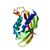
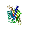
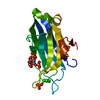
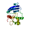
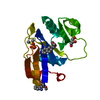
| Title | DDX3 helicase domain |
|---|
 Components Components | ATP-DEPENDENT RNA HELICASE DDX3X |
|---|
 Keywords Keywords | HYDROLASE / PHOSPHORYLATION / NUCLEOTIDE-BINDING / HELICASE / RNA-BINDING / ATP-BINDING / DNA-BINDING / NUCLEAR PROTEIN / HOST-VIRUS INTERACTION |
|---|
| Function / homology |  Function and homology information Function and homology information
CTPase activity / positive regulation of toll-like receptor 8 signaling pathway / positive regulation of toll-like receptor 7 signaling pathway / positive regulation of translation in response to endoplasmic reticulum stress / eukaryotic initiation factor 4E binding / protein localization to cytoplasmic stress granule / RNA strand annealing activity / gamete generation / positive regulation of chemokine (C-C motif) ligand 5 production / positive regulation of mitochondrial translation ...CTPase activity / positive regulation of toll-like receptor 8 signaling pathway / positive regulation of toll-like receptor 7 signaling pathway / positive regulation of translation in response to endoplasmic reticulum stress / eukaryotic initiation factor 4E binding / protein localization to cytoplasmic stress granule / RNA strand annealing activity / gamete generation / positive regulation of chemokine (C-C motif) ligand 5 production / positive regulation of mitochondrial translation / positive regulation of protein K63-linked ubiquitination / cellular response to arsenic-containing substance / NLRP3 inflammasome complex / poly(A) binding / Dengue virus activates/modulates innate and adaptive immune responses / gamma-tubulin binding / P granule / negative regulation of non-canonical NF-kappaB signal transduction / cellular response to osmotic stress / transcription factor binding / negative regulation of intrinsic apoptotic signaling pathway / cytoplasmic pattern recognition receptor signaling pathway / lipid homeostasis / extrinsic apoptotic signaling pathway via death domain receptors / cell leading edge / positive regulation of NLRP3 inflammasome complex assembly / positive regulation of interferon-alpha production / ribosomal small subunit binding / positive regulation of translational initiation / negative regulation of extrinsic apoptotic signaling pathway via death domain receptors / positive regulation of G1/S transition of mitotic cell cycle / positive regulation of viral genome replication / negative regulation of protein-containing complex assembly / positive regulation of type I interferon production / signaling adaptor activity / positive regulation of interferon-beta production / translation initiation factor binding / intrinsic apoptotic signaling pathway / stress granule assembly / DNA helicase activity / protein serine/threonine kinase activator activity / cytosolic ribosome assembly / positive regulation of translation / chromosome segregation / translational initiation / cellular response to virus / negative regulation of cell growth / positive regulation of non-canonical NF-kappaB signal transduction / RNA stem-loop binding / mRNA 5'-UTR binding / Wnt signaling pathway / response to virus / cytoplasmic stress granule / positive regulation of canonical Wnt signaling pathway / ribonucleoside triphosphate phosphatase activity / lamellipodium / positive regulation of cell growth / secretory granule lumen / ficolin-1-rich granule lumen / cell differentiation / RNA helicase activity / negative regulation of translation / intracellular signal transduction / RNA helicase / cadherin binding / positive regulation of apoptotic process / negative regulation of gene expression / innate immune response / mRNA binding / GTPase activity / centrosome / positive regulation of gene expression / Neutrophil degranulation / negative regulation of apoptotic process / DNA-templated transcription / positive regulation of transcription by RNA polymerase II / ATP hydrolysis activity / mitochondrion / DNA binding / RNA binding / extracellular exosome / extracellular region / ATP binding / nucleus / plasma membrane / cytosol / cytoplasmSimilarity search - Function DEAD-box subfamily ATP-dependent helicases signature. / ATP-dependent RNA helicase DEAD-box, conserved site / RNA helicase, DEAD-box type, Q motif / DEAD-box RNA helicase Q motif profile. / DEAD/DEAH box helicase domain / DEAD/DEAH box helicase / Helicase conserved C-terminal domain / helicase superfamily c-terminal domain / Superfamilies 1 and 2 helicase C-terminal domain profile. / Superfamilies 1 and 2 helicase ATP-binding type-1 domain profile. ...DEAD-box subfamily ATP-dependent helicases signature. / ATP-dependent RNA helicase DEAD-box, conserved site / RNA helicase, DEAD-box type, Q motif / DEAD-box RNA helicase Q motif profile. / DEAD/DEAH box helicase domain / DEAD/DEAH box helicase / Helicase conserved C-terminal domain / helicase superfamily c-terminal domain / Superfamilies 1 and 2 helicase C-terminal domain profile. / Superfamilies 1 and 2 helicase ATP-binding type-1 domain profile. / DEAD-like helicases superfamily / Helicase, C-terminal / Helicase superfamily 1/2, ATP-binding domain / P-loop containing nucleotide triphosphate hydrolases / Rossmann fold / P-loop containing nucleoside triphosphate hydrolase / 3-Layer(aba) Sandwich / Alpha BetaSimilarity search - Domain/homology |
|---|
| Biological species |  HOMO SAPIENS (human) HOMO SAPIENS (human) |
|---|
| Method |  X-RAY DIFFRACTION / SYNCHROTRON / MOLECULAR REPLACEMENT / Resolution: 1.91 Å X-RAY DIFFRACTION / SYNCHROTRON / MOLECULAR REPLACEMENT / Resolution: 1.91 Å |
|---|
 Authors Authors | Rodamilans, B. / Montoya, G. |
|---|
 Citation Citation | Journal: Acta Crystallogr.,Sect.F / Year: 2007
Title: Expression, Purification, Crystallization and Preliminary X-Ray Diffraction Analysis of the Ddx3 RNA Helicase Domain.
Authors: Rodamilans, B. / Montoya, G. |
|---|
| History | | Deposition | Feb 13, 2007 | Deposition site: PDBE / Processing site: PDBE |
|---|
| Revision 1.0 | May 13, 2008 | Provider: repository / Type: Initial release |
|---|
| Revision 1.1 | Jul 13, 2011 | Group: Advisory / Version format compliance |
|---|
| Revision 1.2 | May 30, 2012 | Group: Other |
|---|
| Revision 1.3 | Dec 13, 2023 | Group: Data collection / Database references / Refinement description
Category: chem_comp_atom / chem_comp_bond ...chem_comp_atom / chem_comp_bond / database_2 / pdbx_initial_refinement_model
Item: _database_2.pdbx_DOI / _database_2.pdbx_database_accession |
|---|
|
|---|
 Movie
Movie Controller


 Open data
 Basic information
 Structure visualization
 Downloads & links
 Other downloads


 PDBj
PDBj Assembly


 Mass: 18.015 Da / Num. of mol.: 193 / Source method: isolated from a natural source / Formula: H2O
 Sample preparation
 / Beamline: ID23-1 / Wavelength: 0.9
  Processing