 Movie
Movie Controller

[English] 日本語
 Yorodumi
- PDB-3ggn: Crystal structure of DR_A0006 from Deinococcus radiodurans. North... -
+ Open data

- Basic information
| Entry | Database: PDB / ID: 3ggn | ||||||
|---|---|---|---|---|---|---|---|
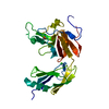
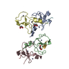
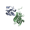
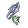
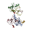
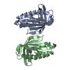
| Title | Crystal structure of DR_A0006 from Deinococcus radiodurans. Northeast Structural Genomics Consortium Target DrR147D | ||||||
 Components Components | Uncharacterized protein DR_A0006 | ||||||
 Keywords Keywords | STRUCTURAL GENOMICS / UNKNOWN FUNCTION / Deinococcus radiodurans / DR_A0006 / PSI-2 / Protein Structure Initiative / Northeast Structural Genomics Consortium / NESG | ||||||
| Function / homology | : / Coenzyme Q-binding protein COQ10, START domain / Polyketide cyclase / dehydrase and lipid transport / START domain / Alpha-D-Glucose-1,6-Bisphosphate; Chain A, domain 4 / START-like domain superfamily / 2-Layer Sandwich / Alpha Beta / Coenzyme Q-binding protein COQ10 START domain-containing protein Function and homology information Function and homology information | ||||||
| Biological species |  Deinococcus radiodurans R1 (radioresistant) Deinococcus radiodurans R1 (radioresistant) | ||||||
| Method |  X-RAY DIFFRACTION / SYNCHROTRON / SAD / Resolution: 2 Å X-RAY DIFFRACTION / SYNCHROTRON / SAD / Resolution: 2 Å | ||||||
 Authors Authors | Seetharaman, J. / Neely, H. / Wang, H. / Janjua, H. / Foote, E.L. / Xiao, R. / Everett, J.K. / Acton, T.B. / Rost, B. / Montelione, G.T. ...Seetharaman, J. / Neely, H. / Wang, H. / Janjua, H. / Foote, E.L. / Xiao, R. / Everett, J.K. / Acton, T.B. / Rost, B. / Montelione, G.T. / Hunt, J.F. / Tong, L. / Northeast Structural Genomics Consortium (NESG) | ||||||
 Citation Citation | Journal: To be Published Title: Crystal structure of DR_A0006 from Deinococcus radiodurans. Authors: Seetharaman, J. / Neely, H. / Wang, H. / Janjua, H. / Foote, E.L. / Xiao, R. / Everett, J.K. / Acton, T.B. / Rost, B. / Montelione, G.T. / Hunt, J.F. / Tong, L. | ||||||
| History |
|
- Structure visualization
| Structure viewer | Molecule: MolmilJmol/JSmol |
|---|
- Downloads & links
-Download
| PDBx/mmCIF format | 3ggn.cif.gz | 64.6 KB | Display | PDBx/mmCIF format |
|---|---|---|---|---|
| PDB format | pdb3ggn.ent.gz | 46.9 KB | Display | PDB format |
| PDBx/mmJSON format | 3ggn.json.gz | Tree view | PDBx/mmJSON format | |
| Others |  Other downloads Other downloads |
-Validation report
| Arichive directory | https://data.pdbj.org/pub/pdb/validation_reports/gg/3ggnftp://data.pdbj.org/pub/pdb/validation_reports/gg/3ggn | HTTPS FTP |
|---|
-Related structure data
| Similar structure data | |
|---|---|
| Other databases |


-Links
 PDBj
PDBj- Assembly
| Deposited unit | 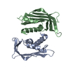
| ||||||||
|---|---|---|---|---|---|---|---|---|---|
| 1 |
| ||||||||
| 2 | 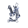
| ||||||||
| 3 | 
| ||||||||
| Unit cell |
|
-Components
| #1: Protein | Mass: 17408.791 Da / Num. of mol.: 2 Source method: isolated from a genetically manipulated source Source: (gene. exp.) Deinococcus radiodurans R1 (radioresistant)Strain: R1 / DSM 20539 / IFO 15346 / LMG 4051 / NCIB 9279 / Gene: DR_A0006 / Plasmid: pET21-23C / Production host: #2: Water | ChemComp-HOH / |  Mass: 18.015 Da / Num. of mol.: 129 / Source method: isolated from a natural source / Formula: H2O Mass: 18.015 Da / Num. of mol.: 129 / Source method: isolated from a natural source / Formula: H2O |
|---|
-Experimental details
-Experiment
| Experiment | Method: X-RAY DIFFRACTION / Number of used crystals: 1 |
|---|
- Sample preparation
| Crystal | Density Matthews: 1.93 Å3/Da / Density % sol: 36.27 % |
|---|---|
| Crystal grow | Temperature: 277 K / Method: microbatch under oil / pH: 6 Details: 0.1M KH2PO4, 0.1M MES pH 6.0, 40% PEG 4000, MICROBATCH UNDER OIL, temperature 277.0K |
-Data collection
| Diffraction | Mean temperature: 100 K |
|---|---|
| Diffraction source | Source: SYNCHROTRON / Site: NSLS  / Beamline: X4A / Wavelength: 0.979 Å / Beamline: X4A / Wavelength: 0.979 Å |
| Detector | Type: ADSC QUANTUM 4 / Detector: CCD / Date: Jan 25, 2009 |
| Radiation | Monochromator: Si(111) / Protocol: SINGLE WAVELENGTH / Monochromatic (M) / Laue (L): M / Scattering type: x-ray |
| Radiation wavelength | Wavelength: 0.979 Å / Relative weight: 1 |
| Reflection | Resolution: 2→50 Å / Num. all: 35348 / Num. obs: 35348 / % possible obs: 98.6 % / Observed criterion σ(F): 0 / Observed criterion σ(I): 0 / Biso Wilson estimate: 10.6 Å2 / Rmerge(I) obs: 0.078 / Rsym value: 0.063 / Net I/σ(I): 14.8 |
| Reflection shell | Resolution: 2→2.07 Å / Rmerge(I) obs: 0.319 / Rsym value: 0.28 / % possible all: 96.4 |
- Processing
| Software |
| ||||||||||||||||||||
|---|---|---|---|---|---|---|---|---|---|---|---|---|---|---|---|---|---|---|---|---|---|
| Refinement | Method to determine structure: SAD / Resolution: 2→37.06 Å / Rfactor Rfree error: 0.007 / Data cutoff high absF: 119679.15 / Data cutoff low absF: 0 / Isotropic thermal model: RESTRAINED / Cross valid method: THROUGHOUT / σ(F): 0 / Stereochemistry target values: Engh & Huber / Details: BULK SOLVENT MODEL USED
| ||||||||||||||||||||
| Solvent computation | Solvent model: FLAT MODEL / Bsol: 59.3031 Å2 / ksol: 0.4 e/Å3 | ||||||||||||||||||||
| Displacement parameters | Biso mean: 26.3 Å2
| ||||||||||||||||||||
| Refine analyze |
| ||||||||||||||||||||
| Refinement step | Cycle: LAST / Resolution: 2→37.06 Å
| ||||||||||||||||||||
| Refine LS restraints |
| ||||||||||||||||||||
| LS refinement shell | Resolution: 2→2.13 Å / Rfactor Rfree error: 0.018 / Total num. of bins used: 6
| ||||||||||||||||||||
| Xplor file |
|
