 Movie
Movie Controller

[English] 日本語
 Yorodumi
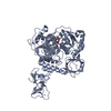
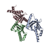
- PDB-3d6r: Structure of an avian influenza A virus NS1 protein effector domain -
+ Open data

- Basic information
| Entry | Database: PDB / ID: 3d6r | ||||||
|---|---|---|---|---|---|---|---|
| Title | Structure of an avian influenza A virus NS1 protein effector domain | ||||||
 Components Components | Non-structural protein 1 | ||||||
 Keywords Keywords | VIRAL PROTEIN / Influenza / NS1 / effector domain / Alternative splicing / Cytoplasm / Host-virus interaction / Interferon antiviral system evasion / Nucleus / RNA-binding / Suppressor of RNA silencing | ||||||
| Function / homology |  Function and homology information Function and homology informationsymbiont-mediated suppression of host mRNA processing / symbiont-mediated suppression of host PKR/eIFalpha signaling / symbiont-mediated suppression of host cytoplasmic pattern recognition receptor signaling pathway via inhibition of RIG-I activity / protein serine/threonine kinase inhibitor activity / host cell cytoplasm / symbiont-mediated suppression of host type I interferon-mediated signaling pathway / symbiont-mediated suppression of host gene expression / host cell nucleus / RNA binding Similarity search - Function | ||||||
| Biological species |   Influenza A virus Influenza A virus | ||||||
| Method |  X-RAY DIFFRACTION / MOLECULAR REPLACEMENT / Resolution: 1.997 Å X-RAY DIFFRACTION / MOLECULAR REPLACEMENT / Resolution: 1.997 Å | ||||||
 Authors Authors | Hale, B.G. / Barclay, W.S. / Randall, R.E. / Russell, R.J. | ||||||
 Citation Citation | Journal: Virology / Year: 2008 Title: Structure of an avian influenza A virus NS1 protein effector domain. Authors: Hale, B.G. / Barclay, W.S. / Randall, R.E. / Russell, R.J. | ||||||
| History |
|
- Structure visualization
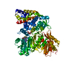
| Structure viewer | Molecule: MolmilJmol/JSmol |
|---|
- Downloads & links
-Download
| PDBx/mmCIF format | 3d6r.cif.gz | 69.5 KB | Display | PDBx/mmCIF format |
|---|---|---|---|---|
| PDB format | pdb3d6r.ent.gz | 51.3 KB | Display | PDB format |
| PDBx/mmJSON format | 3d6r.json.gz | Tree view | PDBx/mmJSON format | |
| Others |  Other downloads Other downloads |
-Validation report
| Arichive directory | https://data.pdbj.org/pub/pdb/validation_reports/d6/3d6rftp://data.pdbj.org/pub/pdb/validation_reports/d6/3d6r | HTTPS FTP |
|---|
-Related structure data
| Similar structure data |
|---|


-Links
 PDBj
PDBj- Assembly
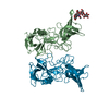
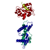
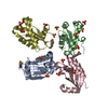
| Deposited unit | 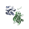
| |||||||||||||||
|---|---|---|---|---|---|---|---|---|---|---|---|---|---|---|---|---|
| 1 | 
| |||||||||||||||
| 2 | 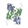
| |||||||||||||||
| 3 | 
| |||||||||||||||
| Unit cell |
| |||||||||||||||
| Components on special symmetry positions |
|
-Components
| #1: Protein | Mass: 17754.348 Da / Num. of mol.: 2 / Fragment: effector domain (UNP residues 73-230) Source method: isolated from a genetically manipulated source Source: (gene. exp.) Influenza A virus / Strain: A/Duck/Alberta/60/1976 H12N5 / Gene: NS / Production host:  #2: Water | ChemComp-HOH / |  Mass: 18.015 Da / Num. of mol.: 387 / Source method: isolated from a natural source / Formula: H2O Mass: 18.015 Da / Num. of mol.: 387 / Source method: isolated from a natural source / Formula: H2O |
|---|
-Experimental details
-Experiment
| Experiment | Method: X-RAY DIFFRACTION / Number of used crystals: 1 |
|---|
- Sample preparation
| Crystal | Density Matthews: 2.93 Å3/Da / Density % sol: 58.04 % |
|---|---|
| Crystal grow | Temperature: 298 K / Method: vapor diffusion, hanging drop / pH: 7 Details: PEG 8K, hepes, Na thiocyanate, glycerol, pH 7.0, VAPOR DIFFUSION, HANGING DROP, temperature 298K |
-Data collection
| Diffraction | Mean temperature: 100 K |
|---|---|
| Diffraction source | Source: ROTATING ANODE / Type: RIGAKU MICROMAX-007 HF / Wavelength: 1.542 Å |
| Detector | Type: RIGAKU SATURN 944 / Detector: CCD / Date: Mar 1, 2008 |
| Radiation | Protocol: SINGLE WAVELENGTH / Monochromatic (M) / Laue (L): M / Scattering type: x-ray |
| Radiation wavelength | Wavelength: 1.542 Å / Relative weight: 1 |
| Reflection | Resolution: 2→30 Å / Num. all: 30438 / Num. obs: 26147 / % possible obs: 85.9 % / Observed criterion σ(F): 0 / Observed criterion σ(I): 0 / Biso Wilson estimate: 24.62 Å2 |
| Reflection shell | Resolution: 2→2.03 Å / % possible all: 49 |
- Processing
| Software |
| |||||||||||||||||||||||||||||||||||||||||||||||||||||||||||||||||||||||||||||
|---|---|---|---|---|---|---|---|---|---|---|---|---|---|---|---|---|---|---|---|---|---|---|---|---|---|---|---|---|---|---|---|---|---|---|---|---|---|---|---|---|---|---|---|---|---|---|---|---|---|---|---|---|---|---|---|---|---|---|---|---|---|---|---|---|---|---|---|---|---|---|---|---|---|---|---|---|---|---|
| Refinement | Method to determine structure: MOLECULAR REPLACEMENT / Resolution: 1.997→19.97 Å / FOM work R set: 0.858 / σ(F): 1.9 / Stereochemistry target values: ML
| |||||||||||||||||||||||||||||||||||||||||||||||||||||||||||||||||||||||||||||
| Solvent computation | Shrinkage radii: 0.9 Å / VDW probe radii: 1.11 Å / Solvent model: FLAT BULK SOLVENT MODEL / Bsol: 58.602 Å2 / ksol: 0.358 e/Å3 | |||||||||||||||||||||||||||||||||||||||||||||||||||||||||||||||||||||||||||||
| Displacement parameters | Biso max: 9.73 Å2 / Biso mean: 26.88 Å2 / Biso min: 74.18 Å2
| |||||||||||||||||||||||||||||||||||||||||||||||||||||||||||||||||||||||||||||
| Refinement step | Cycle: LAST / Resolution: 1.997→19.97 Å
| |||||||||||||||||||||||||||||||||||||||||||||||||||||||||||||||||||||||||||||
| Refine LS restraints |
| |||||||||||||||||||||||||||||||||||||||||||||||||||||||||||||||||||||||||||||
| LS refinement shell | Refine-ID: X-RAY DIFFRACTION / Total num. of bins used: 10
|
