 Movie
Movie Controller

[English] 日本語
 Yorodumi
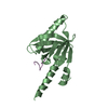
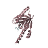
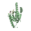
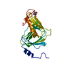
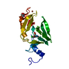
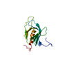
- PDB-1p3r: Crystal structure of the phosphotyrosin binding domain(PTB) of mo... -
+ Open data

- Basic information
| Entry | Database: PDB / ID: 1p3r | ||||||
|---|---|---|---|---|---|---|---|
| Title | Crystal structure of the phosphotyrosin binding domain(PTB) of mouse Disabled 1(Dab1) | ||||||
 Components Components | Disabled homolog 2 | ||||||
 Keywords Keywords | SIGNALING PROTEIN / PTB | ||||||
| Function / homology |  Function and homology information Function and homology informationpositive regulation of aldosterone biosynthetic process / positive regulation of aldosterone secretion / leading edge cell differentiation / pinocytosis / Formation of annular gap junctions / Gap junction degradation / positive regulation of clathrin-dependent endocytosis / positive regulation of early endosome to late endosome transport / renal protein absorption / positive regulation of integrin-mediated signaling pathway ...positive regulation of aldosterone biosynthetic process / positive regulation of aldosterone secretion / leading edge cell differentiation / pinocytosis / Formation of annular gap junctions / Gap junction degradation / positive regulation of clathrin-dependent endocytosis / positive regulation of early endosome to late endosome transport / renal protein absorption / positive regulation of integrin-mediated signaling pathway / AP-2 adaptor complex binding / clathrin coat of coated pit / positive regulation of Wnt signaling pathway, planar cell polarity pathway / Cargo recognition for clathrin-mediated endocytosis / clathrin-coated vesicle membrane / clathrin coat assembly / Clathrin-mediated endocytosis / clathrin-cargo adaptor activity / negative regulation of androgen receptor signaling pathway / endoderm development / myeloid cell differentiation / response to steroid hormone / clathrin-coated vesicle / cargo receptor activity / low-density lipoprotein particle receptor binding / clathrin binding / hematopoietic stem cell proliferation / SMAD binding / positive regulation of receptor recycling / positive regulation of SMAD protein signal transduction / positive regulation of receptor internalization / positive regulation of endocytosis / negative regulation of protein localization to plasma membrane / cellular response to transforming growth factor beta stimulus / positive regulation of epithelial to mesenchymal transition / phosphatidylinositol-4,5-bisphosphate binding / positive regulation of substrate adhesion-dependent cell spreading / clathrin-coated pit / transforming growth factor beta receptor signaling pathway / phosphatidylinositol binding / positive regulation of cell adhesion / receptor-mediated endocytosis / cellular response to epidermal growth factor stimulus / negative regulation of extrinsic apoptotic signaling pathway / positive regulation of transcription elongation by RNA polymerase II / negative regulation of canonical Wnt signaling pathway / negative regulation of cell growth / negative regulation of ERK1 and ERK2 cascade / positive regulation of JNK cascade / integrin binding / cell morphogenesis / fibrillar center / negative regulation of epithelial cell proliferation / Wnt signaling pathway / protein transport / negative regulation of neuron projection development / positive regulation of proteasomal ubiquitin-dependent protein catabolic process / in utero embryonic development / apical plasma membrane / positive regulation of cell migration / apoptotic process / negative regulation of apoptotic process / perinuclear region of cytoplasm / negative regulation of transcription by RNA polymerase II / positive regulation of transcription by RNA polymerase II / nucleus / plasma membrane / cytoplasm Similarity search - Function | ||||||
| Biological species |  | ||||||
| Method |  X-RAY DIFFRACTION / SYNCHROTRON / MOLECULAR REPLACEMENT / Resolution: 2.1 Å X-RAY DIFFRACTION / SYNCHROTRON / MOLECULAR REPLACEMENT / Resolution: 2.1 Å | ||||||
 Authors Authors | Yun, M. / Keshvara, L. / Park, C.G. / Zhang, Y.M. / Dickerson, J.B. / Zheng, J. / Rock, C.O. / Curran, T. / Park, H.W. | ||||||
 Citation Citation | Journal: J.Biol.Chem. / Year: 2003 Title: Crystal structures of the Dab homology domains of mouse disabled 1 and 2. Authors: Yun, M. / Keshvara, L. / Park, C.G. / Zhang, Y.M. / Dickerson, J.B. / Zheng, J. / Rock, C.O. / Curran, T. / Park, H.W. | ||||||
| History |
|
- Structure visualization
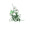
| Structure viewer | Molecule: MolmilJmol/JSmol |
|---|
- Downloads & links
-Download
| PDBx/mmCIF format | 1p3r.cif.gz | 105 KB | Display | PDBx/mmCIF format |
|---|---|---|---|---|
| PDB format | pdb1p3r.ent.gz | 80.8 KB | Display | PDB format |
| PDBx/mmJSON format | 1p3r.json.gz | Tree view | PDBx/mmJSON format | |
| Others |  Other downloads Other downloads |
-Validation report
| Arichive directory | https://data.pdbj.org/pub/pdb/validation_reports/p3/1p3rftp://data.pdbj.org/pub/pdb/validation_reports/p3/1p3r | HTTPS FTP |
|---|
-Related structure data
| Related structure data |  1m7eSC  1oqnC S: Starting model for refinement C: citing same article ( |
|---|---|
| Similar structure data |


-Links
 PDBj
PDBj


- Assembly
| Deposited unit | 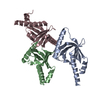
| ||||||||
|---|---|---|---|---|---|---|---|---|---|
| 1 | 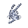
| ||||||||
| 2 | 
| ||||||||
| 3 | 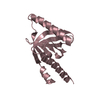
| ||||||||
| Unit cell |
|
-Components
| #1: Protein | Mass: 18042.629 Da / Num. of mol.: 3 / Fragment: PTB domain of mouse Disabled 2 Source method: isolated from a genetically manipulated source Source: (gene. exp.)  #2: Water | ChemComp-HOH / |  Mass: 18.015 Da / Num. of mol.: 315 / Source method: isolated from a natural source / Formula: H2O Mass: 18.015 Da / Num. of mol.: 315 / Source method: isolated from a natural source / Formula: H2O |
|---|
-Experimental details
-Experiment
| Experiment | Method: X-RAY DIFFRACTION / Number of used crystals: 1 |
|---|
- Sample preparation
| Crystal | Density Matthews: 3.97 Å3/Da / Density % sol: 69.06 % | ||||||||||||||||||||||||||||||||||||||||||
|---|---|---|---|---|---|---|---|---|---|---|---|---|---|---|---|---|---|---|---|---|---|---|---|---|---|---|---|---|---|---|---|---|---|---|---|---|---|---|---|---|---|---|---|
| Crystal grow | Temperature: 291 K / Method: vapor diffusion, hanging drop / pH: 8 Details: sodium formate, pH 8.0, VAPOR DIFFUSION, HANGING DROP, temperature 291K | ||||||||||||||||||||||||||||||||||||||||||
| Crystal grow | *PLUS Temperature: 18 ℃ / pH: 7.5 / Method: vapor diffusion, hanging drop | ||||||||||||||||||||||||||||||||||||||||||
| Components of the solutions | *PLUS
|
-Data collection
| Diffraction | Mean temperature: 100 K |
|---|---|
| Diffraction source | Source: SYNCHROTRON / Site: APS  / Beamline: 22-ID / Wavelength: 1.02466 Å / Beamline: 22-ID / Wavelength: 1.02466 Å |
| Detector | Type: MARRESEARCH / Detector: CCD / Date: Dec 8, 2002 |
| Radiation | Protocol: SINGLE WAVELENGTH / Monochromatic (M) / Laue (L): M / Scattering type: x-ray |
| Radiation wavelength | Wavelength: 1.02466 Å / Relative weight: 1 |
| Reflection | Resolution: 2.1→30 Å / Num. obs: 49672 / % possible obs: 97.1 % / Observed criterion σ(I): -1 / Redundancy: 13.5 % / Rsym value: 0.084 / Net I/σ(I): 54.7 |
| Reflection | *PLUS Lowest resolution: 30 Å / Redundancy: 54.7 % / Num. measured all: 2717217 / Rmerge(I) obs: 0.084 |
| Reflection shell | *PLUS Highest resolution: 2.3 Å / Lowest resolution: 2.34 Å / % possible obs: 99.4 % |
- Processing
| Software |
| ||||||||||||||||||
|---|---|---|---|---|---|---|---|---|---|---|---|---|---|---|---|---|---|---|---|
| Refinement | Method to determine structure: MOLECULAR REPLACEMENT Starting model: PDB ENTRY 1M7E Resolution: 2.1→30 Å / σ(F): 1.5 /
| ||||||||||||||||||
| Refinement step | Cycle: LAST / Resolution: 2.1→30 Å
| ||||||||||||||||||
| Refine LS restraints |
| ||||||||||||||||||
| Refinement | *PLUS Lowest resolution: 30 Å / % reflection Rfree: 5 % | ||||||||||||||||||
| Solvent computation | *PLUS | ||||||||||||||||||
| Displacement parameters | *PLUS | ||||||||||||||||||
| Refine LS restraints | *PLUS
|
