 Movie
Movie Controller

[English] 日本語
 Yorodumi
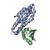
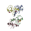
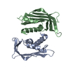
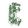
- PDB-6d51: Crystal structure of L,D-transpeptidase 3 from Mycobacterium tube... -
+ Open data

- Basic information
| Entry | Database: PDB / ID: 6d51 | ||||||
|---|---|---|---|---|---|---|---|
| Title | Crystal structure of L,D-transpeptidase 3 from Mycobacterium tuberculosis in complex with a faropenem-derived adduct | ||||||
 Components Components | Probable L,D-transpeptidase 3 | ||||||
 Keywords Keywords | TRANSFERASE / Adduct | ||||||
| Function / homology |  Function and homology information Function and homology informationpeptidoglycan-protein cross-linking / peptidoglycan L,D-transpeptidase activity / Transferases; Acyltransferases; Aminoacyltransferases / acyltransferase activity / cell wall organization / regulation of cell shape Similarity search - Function | ||||||
| Biological species |   Mycobacterium tuberculosis (bacteria) Mycobacterium tuberculosis (bacteria) | ||||||
| Method |  X-RAY DIFFRACTION / SYNCHROTRON / MOLECULAR REPLACEMENT / molecular replacement / Resolution: 1.83 Å X-RAY DIFFRACTION / SYNCHROTRON / MOLECULAR REPLACEMENT / molecular replacement / Resolution: 1.83 Å | ||||||
 Authors Authors | Libreros, G.A. / Dias, M.V.B. | ||||||
| Funding support |  Brazil, 1items Brazil, 1items
| ||||||
 Citation Citation | Journal: ACS Infect Dis / Year: 2019 Title: Structural Basis for the Interaction and Processing of beta-Lactam Antibiotics by l,d-Transpeptidase 3 (LdtMt3) from Mycobacterium tuberculosis. Authors: Libreros-Zuniga, G.A. / Dos Santos Silva, C. / Salgado Ferreira, R. / Dias, M.V.B. | ||||||
| History |
|
- Structure visualization
| Structure viewer | Molecule: MolmilJmol/JSmol |
|---|
- Downloads & links
-Download
| PDBx/mmCIF format | 6d51.cif.gz | 67 KB | Display | PDBx/mmCIF format |
|---|---|---|---|---|
| PDB format | pdb6d51.ent.gz | 45.6 KB | Display | PDB format |
| PDBx/mmJSON format | 6d51.json.gz | Tree view | PDBx/mmJSON format | |
| Others |  Other downloads Other downloads |
-Validation report
| Arichive directory | https://data.pdbj.org/pub/pdb/validation_reports/d5/6d51ftp://data.pdbj.org/pub/pdb/validation_reports/d5/6d51 | HTTPS FTP |
|---|
-Related structure data
| Related structure data | 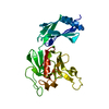 6d4kC  6d5aC  4k73S S: Starting model for refinement C: citing same article ( |
|---|---|
| Similar structure data |


-Links
 PDBj
PDBj- Assembly
| Deposited unit | 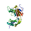
| ||||||||
|---|---|---|---|---|---|---|---|---|---|
| 1 |
| ||||||||
| Unit cell |
|
-Components
| #1: Protein | Mass: 27953.246 Da / Num. of mol.: 1 / Fragment: residues 33-271 Source method: isolated from a genetically manipulated source Source: (gene. exp.) Mycobacterium tuberculosis (bacteria) / Strain: ATCC 25618 / H37Rv / Gene: Rv1433, RVBD_1433, P425_01489 / Production host: References: UniProt: O06825, Transferases; Acyltransferases; Aminoacyltransferases |
|---|---|
| #2: Chemical | ChemComp-ACE /   Mass: 44.053 Da / Num. of mol.: 1 / Source method: obtained synthetically / Formula: C2H4O Mass: 44.053 Da / Num. of mol.: 1 / Source method: obtained synthetically / Formula: C2H4O |
| #3: Chemical | ChemComp-CA /   Mass: 40.078 Da / Num. of mol.: 1 / Source method: obtained synthetically / Formula: Ca Mass: 40.078 Da / Num. of mol.: 1 / Source method: obtained synthetically / Formula: Ca |
| #4: Water | ChemComp-HOH /  Mass: 18.015 Da / Num. of mol.: 199 / Source method: isolated from a natural source / Formula: H2O Mass: 18.015 Da / Num. of mol.: 199 / Source method: isolated from a natural source / Formula: H2O |
| Nonpolymer details | THE AUTHORS STATE THAT SOME BETA LACTAM ANTIBIOTICS, SUCH AS FAROPENEM, CAN BE PROCESSED OR ...THE AUTHORS STATE THAT SOME BETA LACTAM ANTIBIOTIC |
-Experimental details
-Experiment
| Experiment | Method: X-RAY DIFFRACTION / Number of used crystals: 1 |
|---|
- Sample preparation
| Crystal | Density Matthews: 2.2 Å3/Da / Density % sol: 38.96 % |
|---|---|
| Crystal grow | Temperature: 291 K / Method: vapor diffusion, hanging drop / pH: 7.5 Details: PEG 8000 10% (w/v), Hepes 100 mM, calcium acetate 200 mM |
-Data collection
| Diffraction | Mean temperature: 100 K | ||||||||||||||||||||||||||||||
|---|---|---|---|---|---|---|---|---|---|---|---|---|---|---|---|---|---|---|---|---|---|---|---|---|---|---|---|---|---|---|---|
| Diffraction source | Source: SYNCHROTRON / Site: LNLS / Beamline: W01B-MX2 / Wavelength: 1.458 Å | ||||||||||||||||||||||||||||||
| Detector | Type: DECTRIS PILATUS 2M / Detector: PIXEL / Date: May 30, 2017 | ||||||||||||||||||||||||||||||
| Radiation | Protocol: SINGLE WAVELENGTH / Monochromatic (M) / Laue (L): M / Scattering type: x-ray | ||||||||||||||||||||||||||||||
| Radiation wavelength | Wavelength: 1.458 Å / Relative weight: 1 | ||||||||||||||||||||||||||||||
| Reflection | Resolution: 1.83→46.17 Å / Num. obs: 20633 / % possible obs: 99.9 % / Redundancy: 12.3 % / Biso Wilson estimate: 26.58 Å2 / CC1/2: 0.999 / Rmerge(I) obs: 0.076 / Rpim(I) all: 0.023 / Rrim(I) all: 0.079 / Net I/σ(I): 20.4 / Num. measured all: 253996 / Scaling rejects: 6 | ||||||||||||||||||||||||||||||
| Reflection shell | Diffraction-ID: 1
|
-Phasing
| Phasing | Method: molecular replacement |
|---|
- Processing
| Software |
| ||||||||||||||||||||||||||||||||||||||||||||||||||||||||
|---|---|---|---|---|---|---|---|---|---|---|---|---|---|---|---|---|---|---|---|---|---|---|---|---|---|---|---|---|---|---|---|---|---|---|---|---|---|---|---|---|---|---|---|---|---|---|---|---|---|---|---|---|---|---|---|---|---|
| Refinement | Method to determine structure: MOLECULAR REPLACEMENT Starting model: 4K73 Resolution: 1.83→40.944 Å / SU ML: 0.25 / Cross valid method: THROUGHOUT / σ(F): 1.35 / Phase error: 25.19
| ||||||||||||||||||||||||||||||||||||||||||||||||||||||||
| Solvent computation | Shrinkage radii: 0.9 Å / VDW probe radii: 1.11 Å | ||||||||||||||||||||||||||||||||||||||||||||||||||||||||
| Displacement parameters | Biso max: 99.85 Å2 / Biso mean: 37.8167 Å2 / Biso min: 14.88 Å2 | ||||||||||||||||||||||||||||||||||||||||||||||||||||||||
| Refinement step | Cycle: final / Resolution: 1.83→40.944 Å
| ||||||||||||||||||||||||||||||||||||||||||||||||||||||||
| Refine LS restraints |
| ||||||||||||||||||||||||||||||||||||||||||||||||||||||||
| LS refinement shell | Refine-ID: X-RAY DIFFRACTION / Rfactor Rfree error: 0 / Total num. of bins used: 7
|
