| Entry | Database: PDB / ID: 5by3
|
|---|
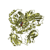
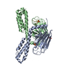
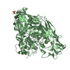
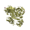
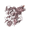
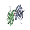
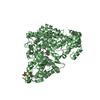
| Title | A novel family GH115 4-O-Methyl-alpha-glucuronidase, BtGH115A, with specificity for decorated arabinogalactans |
|---|
 Components Components | BtGH115A |
|---|
 Keywords Keywords | SUGAR BINDING PROTEIN / Glycoside / hydrolase / arabinogalactans alpha-glucuronidase |
|---|
| Function / homology |  Function and homology information Function and homology information
Glycosyl hydrolase family 115 / Glycosyl hydrolase family 115 / Gylcosyl hydrolase 115 C-terminal domain / Glycosyl hydrolase 115 superfamily / Glycosyl hydrolase family 115 / Glycosyl hydrolase family 115 C-terminal domain / Chitobiase/beta-hexosaminidase domain 2-like / Chitobiase; domain 2 / Beta-hexosaminidase-like, domain 2 / TIM Barrel ...Glycosyl hydrolase family 115 / Glycosyl hydrolase family 115 / Gylcosyl hydrolase 115 C-terminal domain / Glycosyl hydrolase 115 superfamily / Glycosyl hydrolase family 115 / Glycosyl hydrolase family 115 C-terminal domain / Chitobiase/beta-hexosaminidase domain 2-like / Chitobiase; domain 2 / Beta-hexosaminidase-like, domain 2 / TIM Barrel / Alpha-Beta Barrel / 2-Layer Sandwich / Alpha BetaSimilarity search - Domain/homology |
|---|
| Biological species |  Bacteroides thetaiotaomicron (bacteria) Bacteroides thetaiotaomicron (bacteria) |
|---|
| Method |  X-RAY DIFFRACTION / SYNCHROTRON / Resolution: 2.44 Å X-RAY DIFFRACTION / SYNCHROTRON / Resolution: 2.44 Å |
|---|
 Authors Authors | Lammerts van Bueren, A. / Davies, G.J. / Turkenburg, J.P. |
|---|
 Citation Citation | Journal: J.Mol.Biol. / Year: 2015
Title: Structural and Functional Characterization of a Novel Family GH115 4-O-Methyl-alpha-Glucuronidase with Specificity for Decorated Arabinogalactans.
Authors: Aalbers, F. / Turkenburg, J.P. / Davies, G.J. / Dijkhuizen, L. / Lammerts van Bueren, A. |
|---|
| History | | Deposition | Jun 10, 2015 | Deposition site: RCSB / Processing site: PDBE |
|---|
| Revision 1.0 | Jul 22, 2015 | Provider: repository / Type: Initial release |
|---|
| Revision 1.1 | Aug 5, 2015 | Group: Database references |
|---|
| Revision 1.2 | Dec 9, 2015 | Group: Database references |
|---|
| Revision 1.3 | Nov 13, 2024 | Group: Data collection / Database references ...Data collection / Database references / Derived calculations / Structure summary
Category: chem_comp_atom / chem_comp_bond ...chem_comp_atom / chem_comp_bond / database_2 / pdbx_entry_details / pdbx_modification_feature / pdbx_struct_conn_angle / struct_conn
Item: _database_2.pdbx_DOI / _database_2.pdbx_database_accession ..._database_2.pdbx_DOI / _database_2.pdbx_database_accession / _pdbx_struct_conn_angle.ptnr1_auth_seq_id / _pdbx_struct_conn_angle.ptnr1_label_alt_id / _pdbx_struct_conn_angle.ptnr3_auth_seq_id / _pdbx_struct_conn_angle.ptnr3_label_alt_id / _pdbx_struct_conn_angle.value / _struct_conn.conn_type_id / _struct_conn.id / _struct_conn.pdbx_dist_value / _struct_conn.pdbx_leaving_atom_flag / _struct_conn.pdbx_ptnr2_label_alt_id / _struct_conn.ptnr1_auth_comp_id / _struct_conn.ptnr1_auth_seq_id / _struct_conn.ptnr1_label_atom_id / _struct_conn.ptnr1_label_comp_id / _struct_conn.ptnr1_label_seq_id / _struct_conn.ptnr2_auth_comp_id / _struct_conn.ptnr2_auth_seq_id / _struct_conn.ptnr2_label_asym_id / _struct_conn.ptnr2_label_atom_id / _struct_conn.ptnr2_label_comp_id / _struct_conn.ptnr2_label_seq_id |
|---|
|
|---|
 Movie
Movie Controller

 Yorodumi
 Open data
 Basic information
 Structure visualization
 Downloads & links
 Other downloads
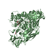


 PDBj
PDBj
 Assembly

 Mass: 96.063 Da / Num. of mol.: 1 / Source method: obtained synthetically / Formula: SO4
 Mass: 58.693 Da / Num. of mol.: 1 / Source method: obtained synthetically / Formula: Ni
 Mass: 18.015 Da / Num. of mol.: 99 / Source method: isolated from a natural source / Formula: H2O
 Sample preparation
 / Beamline: I02 / Wavelength: 0.9795, 0.9798, 0.9682
 Processing