 Movie
Movie Controller

[English] 日本語
 Yorodumi
- PDB-5bu8: HK620 Tail Needle crystallized at pH 7.5 and derivatized with Xenon -
+ Open data

- Basic information
| Entry | Database: PDB / ID: 5bu8 | ||||||
|---|---|---|---|---|---|---|---|
| Title | HK620 Tail Needle crystallized at pH 7.5 and derivatized with Xenon | ||||||
 Components Components | (DNA stabilization protein) x 2 | ||||||
 Keywords Keywords | VIRAL PROTEIN / HK620 / Tail Needle / Membrane penetration | ||||||
| Function / homology |  Function and homology information Function and homology informationsymbiont genome ejection through host cell envelope, short tail mechanism / virus tail / symbiont genome entry into host cell via pore formation in plasma membrane / metal ion binding Similarity search - Function | ||||||
| Biological species |  Salmonella phage HK620 (virus) Salmonella phage HK620 (virus) | ||||||
| Method |  X-RAY DIFFRACTION / SYNCHROTRON / MOLECULAR REPLACEMENT / Resolution: 2.202 Å X-RAY DIFFRACTION / SYNCHROTRON / MOLECULAR REPLACEMENT / Resolution: 2.202 Å | ||||||
 Authors Authors | Sankhala, R.S. / Cingolani, G. | ||||||
| Funding support |  United States, 1items United States, 1items
| ||||||
 Citation Citation | Journal: J.Biol.Chem. / Year: 2016 Title: Structural Plasticity of the Protein Plug That Traps Newly Packaged Genomes in Podoviridae Virions. Authors: Bhardwaj, A. / Sankhala, R.S. / Olia, A.S. / Brooke, D. / Casjens, S.R. / Taylor, D.J. / Prevelige, P.E. / Cingolani, G. | ||||||
| History |
|
- Structure visualization
| Structure viewer | Molecule: MolmilJmol/JSmol |
|---|
- Downloads & links
-Download
| PDBx/mmCIF format | 5bu8.cif.gz | 157.7 KB | Display | PDBx/mmCIF format |
|---|---|---|---|---|
| PDB format | pdb5bu8.ent.gz | 121.8 KB | Display | PDB format |
| PDBx/mmJSON format | 5bu8.json.gz | Tree view | PDBx/mmJSON format | |
| Others |  Other downloads Other downloads |
-Validation report
| Arichive directory | https://data.pdbj.org/pub/pdb/validation_reports/bu/5bu8ftp://data.pdbj.org/pub/pdb/validation_reports/bu/5bu8 | HTTPS FTP |
|---|
-Related structure data
| Related structure data | 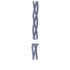 4zkpC 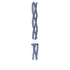 4zkuC 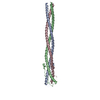 4zxqC  5bu5C  5bvzC  2pohS C: citing same article ( S: Starting model for refinement |
|---|---|
| Similar structure data |


-Links
 PDBj
PDBj
- Assembly
| Deposited unit | 
| ||||||||||||||||||||||||||||||||||||
|---|---|---|---|---|---|---|---|---|---|---|---|---|---|---|---|---|---|---|---|---|---|---|---|---|---|---|---|---|---|---|---|---|---|---|---|---|---|
| 1 | 
| ||||||||||||||||||||||||||||||||||||
| 2 | 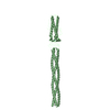
| ||||||||||||||||||||||||||||||||||||
| Unit cell |
| ||||||||||||||||||||||||||||||||||||
| Components on special symmetry positions |
|
-Components
| #1: Protein | Mass: 20994.432 Da / Num. of mol.: 1 Source method: isolated from a genetically manipulated source Source: (gene. exp.) Salmonella phage HK620 (virus) / Gene: 26 / Production host:  | ||||||||
|---|---|---|---|---|---|---|---|---|---|
| #2: Protein | Mass: 25084.785 Da / Num. of mol.: 1 Source method: isolated from a genetically manipulated source Source: (gene. exp.) Salmonella phage HK620 (virus) / Gene: 26 / Production host: | ||||||||
| #3: Chemical |   Mass: 40.078 Da / Num. of mol.: 2 / Source method: obtained synthetically / Formula: Ca Mass: 40.078 Da / Num. of mol.: 2 / Source method: obtained synthetically / Formula: Ca#4: Chemical | ChemComp-XE /   Mass: 131.293 Da / Num. of mol.: 6 / Source method: obtained synthetically / Formula: Xe Mass: 131.293 Da / Num. of mol.: 6 / Source method: obtained synthetically / Formula: Xe#5: Water | ChemComp-HOH / |  Mass: 18.015 Da / Num. of mol.: 300 / Source method: isolated from a natural source / Formula: H2O Mass: 18.015 Da / Num. of mol.: 300 / Source method: isolated from a natural source / Formula: H2OHas protein modification | N | Sequence details | Residues 26-46 in chain A had been modeled as a poly ALA but represented here as UNK residues in ...Residues 26-46 in chain A had been modeled as a poly ALA but represented here as UNK residues in the coordinates. Residue numbers are arbitrary as the exact registry is not clear. Full sequence is the same as that of the chain B. | |
-Experimental details
-Experiment
| Experiment | Method: X-RAY DIFFRACTION / Number of used crystals: 1 |
|---|
- Sample preparation
| Crystal | Density Matthews: 2.97 Å3/Da / Density % sol: 58.59 % |
|---|---|
| Crystal grow | Temperature: 293 K / Method: vapor diffusion, hanging drop / pH: 7.5 Details: 30% PEG 1000, 0.1M Tris pH 7.5 derivatized with Xenon gas prior to data collection PH range: 7.5 |
-Data collection
| Diffraction | Mean temperature: 103 K |
|---|---|
| Diffraction source | Source: SYNCHROTRON / Site: CHESS / Beamline: F1 / Wavelength: 0.92 Å |
| Detector | Type: ADSC QUANTUM 270 / Detector: CCD / Date: May 6, 2007 |
| Radiation | Monochromator: SI 111 / Protocol: SINGLE WAVELENGTH / Monochromatic (M) / Laue (L): M / Scattering type: x-ray |
| Radiation wavelength | Wavelength: 0.92 Å / Relative weight: 1 |
| Reflection | Resolution: 2.2→28.988 Å / Num. obs: 26538 / % possible obs: 90.5 % / Redundancy: 4.2 % / Biso Wilson estimate: 12.7 Å2 / Rmerge(I) obs: 0.1 / Rsym value: 0.11 / Net I/σ(I): 23.5 |
| Reflection shell | Resolution: 2.2→2.28 Å / Redundancy: 1.9 % / Mean I/σ(I) obs: 7.9 / % possible all: 58.5 |
- Processing
| Software |
| |||||||||||||||||||||||||||||||||||||||||||||||||||||||||||||||||||||||||||||||||||||||||||||||||||||||||||||||||||||||||||||
|---|---|---|---|---|---|---|---|---|---|---|---|---|---|---|---|---|---|---|---|---|---|---|---|---|---|---|---|---|---|---|---|---|---|---|---|---|---|---|---|---|---|---|---|---|---|---|---|---|---|---|---|---|---|---|---|---|---|---|---|---|---|---|---|---|---|---|---|---|---|---|---|---|---|---|---|---|---|---|---|---|---|---|---|---|---|---|---|---|---|---|---|---|---|---|---|---|---|---|---|---|---|---|---|---|---|---|---|---|---|---|---|---|---|---|---|---|---|---|---|---|---|---|---|---|---|---|
| Refinement | Method to determine structure: MOLECULAR REPLACEMENT Starting model: 2POH Resolution: 2.202→28.988 Å / Cross valid method: FREE R-VALUE / σ(F): 0.25 / Phase error: 26.72 / Stereochemistry target values: TWIN_LSQ_F
| |||||||||||||||||||||||||||||||||||||||||||||||||||||||||||||||||||||||||||||||||||||||||||||||||||||||||||||||||||||||||||||
| Solvent computation | Shrinkage radii: 0.9 Å / VDW probe radii: 1.11 Å / Solvent model: FLAT BULK SOLVENT MODEL | |||||||||||||||||||||||||||||||||||||||||||||||||||||||||||||||||||||||||||||||||||||||||||||||||||||||||||||||||||||||||||||
| Refinement step | Cycle: LAST / Resolution: 2.202→28.988 Å
| |||||||||||||||||||||||||||||||||||||||||||||||||||||||||||||||||||||||||||||||||||||||||||||||||||||||||||||||||||||||||||||
| Refine LS restraints |
| |||||||||||||||||||||||||||||||||||||||||||||||||||||||||||||||||||||||||||||||||||||||||||||||||||||||||||||||||||||||||||||
| LS refinement shell |
| |||||||||||||||||||||||||||||||||||||||||||||||||||||||||||||||||||||||||||||||||||||||||||||||||||||||||||||||||||||||||||||
| Refinement TLS params. | Method: refined / Refine-ID: X-RAY DIFFRACTION
| |||||||||||||||||||||||||||||||||||||||||||||||||||||||||||||||||||||||||||||||||||||||||||||||||||||||||||||||||||||||||||||
| Refinement TLS group |
|
