 Movie
Movie Controller

[English] 日本語
 Yorodumi
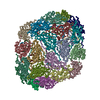
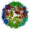
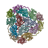
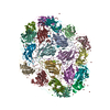
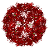
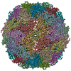
- PDB-5bkq: Crystallographic structure of a cubic form of STMV grown from nitrate -
+ Open data

- Basic information
| Entry | Database: PDB / ID: 5bkq | ||||||
|---|---|---|---|---|---|---|---|
| Title | Crystallographic structure of a cubic form of STMV grown from nitrate | ||||||
 Components Components | Coat protein | ||||||
 Keywords Keywords | VIRUS / ions / RNA / decapsidation / ion channel / crystallization | ||||||
| Function / homology | Satellite virus coat domain superfamily / viral capsid / structural molecule activity / NITRATE ION / Coat protein Function and homology information Function and homology information | ||||||
| Biological species |  Satellite tobacco mosaic virus Satellite tobacco mosaic virus | ||||||
| Method |  X-RAY DIFFRACTION / SYNCHROTRON / MOLECULAR REPLACEMENT / Resolution: 3.19 Å X-RAY DIFFRACTION / SYNCHROTRON / MOLECULAR REPLACEMENT / Resolution: 3.19 Å | ||||||
 Authors Authors | McPherson, A. | ||||||
 Citation Citation | Journal: Acta Crystallogr.,Sect.F / Year: 2021 Title: Structures of additional crystal forms of Satellite tobacco mosaic virus grown from a variety of salts. Authors: McPherson, A. | ||||||
| History |
|
- Structure visualization
| Structure viewer | Molecule: MolmilJmol/JSmol |
|---|
- Downloads & links
-Download
| PDBx/mmCIF format | 5bkq.cif.gz | 2 MB | Display | PDBx/mmCIF format |
|---|---|---|---|---|
| PDB format | pdb5bkq.ent.gz | Display | PDB format | |
| PDBx/mmJSON format | 5bkq.json.gz | Tree view | PDBx/mmJSON format | |
| Others |  Other downloads Other downloads |
-Validation report
| Arichive directory | https://data.pdbj.org/pub/pdb/validation_reports/bk/5bkqftp://data.pdbj.org/pub/pdb/validation_reports/bk/5bkq | HTTPS FTP |
|---|
-Related structure data
| Related structure data | 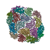 5bklC 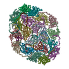 5bknC 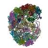 7m2tC  7m2vC 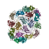 7m3rC 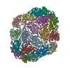 7m3tC 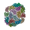 7m50C  7m54C 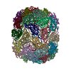 7m57C  4oq8S S: Starting model for refinement C: citing same article ( |
|---|---|
| Similar structure data |


-Links
 PDBj
PDBj
- Assembly
| Deposited unit | 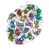
| |||||||||||||||||||||||||||||||||||||||||||||||||||||||||||||||
|---|---|---|---|---|---|---|---|---|---|---|---|---|---|---|---|---|---|---|---|---|---|---|---|---|---|---|---|---|---|---|---|---|---|---|---|---|---|---|---|---|---|---|---|---|---|---|---|---|---|---|---|---|---|---|---|---|---|---|---|---|---|---|---|---|
| 1 | 
| |||||||||||||||||||||||||||||||||||||||||||||||||||||||||||||||
| Unit cell |
| |||||||||||||||||||||||||||||||||||||||||||||||||||||||||||||||
| Components on special symmetry positions |
|
-Components
| #1: Protein | Mass: 17533.949 Da / Num. of mol.: 20 / Source method: isolated from a natural source / Source: (natural) Satellite tobacco mosaic virus / References: UniProt: P17574#2: Chemical | ChemComp-NO3 /   Mass: 62.005 Da / Num. of mol.: 102 / Source method: obtained synthetically / Formula: NO3 / Feature type: SUBJECT OF INVESTIGATION Mass: 62.005 Da / Num. of mol.: 102 / Source method: obtained synthetically / Formula: NO3 / Feature type: SUBJECT OF INVESTIGATION#3: Chemical | ChemComp-MG /   Mass: 24.305 Da / Num. of mol.: 9 / Source method: obtained synthetically / Formula: Mg Mass: 24.305 Da / Num. of mol.: 9 / Source method: obtained synthetically / Formula: Mg#4: Water | ChemComp-HOH / |  Mass: 18.015 Da / Num. of mol.: 1583 / Source method: isolated from a natural source / Formula: H2O Mass: 18.015 Da / Num. of mol.: 1583 / Source method: isolated from a natural source / Formula: H2OHas ligand of interest | Y | |
|---|
-Experimental details
-Experiment
| Experiment | Method: X-RAY DIFFRACTION / Number of used crystals: 1 |
|---|
- Sample preparation
| Crystal | Density Matthews: 3.05 Å3/Da / Density % sol: 59.63 % / Description: octahedra |
|---|---|
| Crystal grow | Temperature: 279 K / Method: vapor diffusion, sitting drop / pH: 6 Details: Crystals were grown by sitting drop vapor diffusion in Cryschem plates using 0.6 ml reservoirs. Drops were equal amounts of a 5 mg/ml virus stock solution buffered with 0.1 M phosphate at pH ...Details: Crystals were grown by sitting drop vapor diffusion in Cryschem plates using 0.6 ml reservoirs. Drops were equal amounts of a 5 mg/ml virus stock solution buffered with 0.1 M phosphate at pH 6.5, and the reservoir solution which was 20% w/v sodium nitrate in 0.1 M phosphate at pH 6.0. Crystallization was carried out at 4 degrees C. PH range: 4.5 - 7.0 |
-Data collection
| Diffraction | Mean temperature: 173 K / Serial crystal experiment: N |
|---|---|
| Diffraction source | Source: SYNCHROTRON / Site: ALS  / Beamline: 8.3.1 / Wavelength: 1 Å / Beamline: 8.3.1 / Wavelength: 1 Å |
| Detector | Type: MAR CCD 165 mm / Detector: CCD / Date: Jun 19, 2002 |
| Radiation | Protocol: SINGLE WAVELENGTH / Monochromatic (M) / Laue (L): M / Scattering type: x-ray |
| Radiation wavelength | Wavelength: 1 Å / Relative weight: 1 |
| Reflection | Resolution: 3.18→105 Å / Num. obs: 45796 / % possible obs: 0.65 % / Redundancy: 3.8 % / Biso Wilson estimate: 50.45 Å2 / CC1/2: 0.964 / Rmerge(I) obs: 0.143 / Rsym value: 0.142 / Net I/σ(I): 11.5 |
| Reflection shell | Resolution: 3.18→3.25 Å / Rmerge(I) obs: 0.235 / Mean I/σ(I) obs: 2.2 / Num. unique obs: 6489 / CC1/2: 0.46 |
- Processing
| Software |
| |||||||||||||||||||||||||||||||||||||||||||||||||||||||||||||||||||||||||||||||||||||||||||||||||||||||||||||||||||||||
|---|---|---|---|---|---|---|---|---|---|---|---|---|---|---|---|---|---|---|---|---|---|---|---|---|---|---|---|---|---|---|---|---|---|---|---|---|---|---|---|---|---|---|---|---|---|---|---|---|---|---|---|---|---|---|---|---|---|---|---|---|---|---|---|---|---|---|---|---|---|---|---|---|---|---|---|---|---|---|---|---|---|---|---|---|---|---|---|---|---|---|---|---|---|---|---|---|---|---|---|---|---|---|---|---|---|---|---|---|---|---|---|---|---|---|---|---|---|---|---|---|
| Refinement | Method to determine structure: MOLECULAR REPLACEMENT Starting model: 4OQ8 Resolution: 3.19→75 Å / SU ML: 0.459 / Cross valid method: FREE R-VALUE / σ(F): 1.33 / Phase error: 33.7232 Stereochemistry target values: GeoStd + Monomer Library + CDL v1.2
| |||||||||||||||||||||||||||||||||||||||||||||||||||||||||||||||||||||||||||||||||||||||||||||||||||||||||||||||||||||||
| Solvent computation | Shrinkage radii: 0.9 Å / VDW probe radii: 1.11 Å / Solvent model: FLAT BULK SOLVENT MODEL | |||||||||||||||||||||||||||||||||||||||||||||||||||||||||||||||||||||||||||||||||||||||||||||||||||||||||||||||||||||||
| Displacement parameters | Biso mean: 54.84 Å2 | |||||||||||||||||||||||||||||||||||||||||||||||||||||||||||||||||||||||||||||||||||||||||||||||||||||||||||||||||||||||
| Refinement step | Cycle: LAST / Resolution: 3.19→75 Å
| |||||||||||||||||||||||||||||||||||||||||||||||||||||||||||||||||||||||||||||||||||||||||||||||||||||||||||||||||||||||
| Refine LS restraints |
| |||||||||||||||||||||||||||||||||||||||||||||||||||||||||||||||||||||||||||||||||||||||||||||||||||||||||||||||||||||||
| LS refinement shell |
| |||||||||||||||||||||||||||||||||||||||||||||||||||||||||||||||||||||||||||||||||||||||||||||||||||||||||||||||||||||||
| Refinement TLS params. | Method: refined / Origin x: 90.5462985363 Å / Origin y: 176.128540856 Å / Origin z: 29.0504143514 Å
| |||||||||||||||||||||||||||||||||||||||||||||||||||||||||||||||||||||||||||||||||||||||||||||||||||||||||||||||||||||||
| Refinement TLS group | Selection details: all |
