 Movie
Movie Controller

[English] 日本語
 Yorodumi
- PDB-5b3g: The crystal structure of the heterodimer of SHORT-ROOT and SCAREC... -
+ Open data

- Basic information
| Entry | Database: PDB / ID: 5b3g | |||||||||
|---|---|---|---|---|---|---|---|---|---|---|
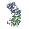
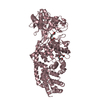
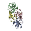
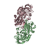
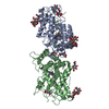
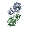
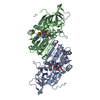
| Title | The crystal structure of the heterodimer of SHORT-ROOT and SCARECROW GRAS domains | |||||||||
 Components Components |
| |||||||||
 Keywords Keywords | TRANSCRIPTION / Transcription cofactor | |||||||||
| Function / homology |  Function and homology information Function and homology informationbundle sheath cell fate specification / radial pattern formation / regulation of hormone metabolic process / gravitropism / asymmetric cell division / leaf development / root development / negative regulation of mitotic cell cycle / maintenance of protein location in nucleus / cell redox homeostasis ...bundle sheath cell fate specification / radial pattern formation / regulation of hormone metabolic process / gravitropism / asymmetric cell division / leaf development / root development / negative regulation of mitotic cell cycle / maintenance of protein location in nucleus / cell redox homeostasis / multicellular organismal-level iron ion homeostasis / recycling endosome / late endosome / sequence-specific DNA binding / early endosome / transcription cis-regulatory region binding / DNA-binding transcription factor activity / regulation of DNA-templated transcription / DNA-templated transcription / nucleus Similarity search - Function | |||||||||
| Biological species |  | |||||||||
| Method |  X-RAY DIFFRACTION / SYNCHROTRON / SAD / Resolution: 2 Å X-RAY DIFFRACTION / SYNCHROTRON / SAD / Resolution: 2 Å | |||||||||
 Authors Authors | Hirano, Y. / Nakagawa, M. / Hakoshima, T. | |||||||||
| Funding support |  Japan, 2items Japan, 2items
| |||||||||
 Citation Citation | Journal: Nat Plants / Year: 2017 Title: Structure of the SHR-SCR heterodimer bound to the BIRD/IDD transcriptional factor JKD Authors: Hirano, Y. / Nakagawa, M. / Suyama, T. / Murase, K. / Shirakawa, M. / Takayama, S. / Sun, T.P. / Hakoshima, T. | |||||||||
| History |
|
- Structure visualization
| Structure viewer | Molecule: MolmilJmol/JSmol |
|---|
- Downloads & links
-Download
| PDBx/mmCIF format | 5b3g.cif.gz | 175.6 KB | Display | PDBx/mmCIF format |
|---|---|---|---|---|
| PDB format | pdb5b3g.ent.gz | 134.1 KB | Display | PDB format |
| PDBx/mmJSON format | 5b3g.json.gz | Tree view | PDBx/mmJSON format | |
| Others |  Other downloads Other downloads |
-Validation report
| Arichive directory | https://data.pdbj.org/pub/pdb/validation_reports/b3/5b3gftp://data.pdbj.org/pub/pdb/validation_reports/b3/5b3g | HTTPS FTP |
|---|
-Related structure data


-Links
 PDBj
PDBj- Assembly
| Deposited unit | 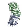
| ||||||||
|---|---|---|---|---|---|---|---|---|---|
| 1 |
| ||||||||
| Unit cell |
|
-Components
-Protein , 2 types, 2 molecules AB
| #1: Protein | Mass: 42212.922 Da / Num. of mol.: 1 / Fragment: UNP residues 274-653 Source method: isolated from a genetically manipulated source Source: (gene. exp.)  |
|---|---|
| #2: Protein | Mass: 53035.047 Da / Num. of mol.: 1 / Fragment: UNP residues 59-531 Source method: isolated from a genetically manipulated source Source: (gene. exp.) |
-Non-polymers , 4 types, 293 molecules 


| #3: Chemical | ChemComp-PO4 /  Mass: 94.971 Da / Num. of mol.: 1 / Source method: obtained synthetically / Formula: PO4 Mass: 94.971 Da / Num. of mol.: 1 / Source method: obtained synthetically / Formula: PO4 | ||||
|---|---|---|---|---|---|
| #4: Chemical |  Mass: 62.068 Da / Num. of mol.: 3 / Source method: obtained synthetically / Formula: C2H6O2 Mass: 62.068 Da / Num. of mol.: 3 / Source method: obtained synthetically / Formula: C2H6O2#5: Chemical |  Mass: 106.120 Da / Num. of mol.: 3 / Source method: obtained synthetically / Formula: C4H10O3 Mass: 106.120 Da / Num. of mol.: 3 / Source method: obtained synthetically / Formula: C4H10O3#6: Water | ChemComp-HOH / | Mass: 18.015 Da / Num. of mol.: 286 / Source method: isolated from a natural source / Formula: H2O |
-Details
| Sequence details | SEQUENCE CONFLICT P233S IS BASED ON REFERENCE 4 (AAL69513) ACCORDING TO DATABASE Q9SZF7 (SHR_ARATH) |
|---|
-Experimental details
-Experiment
| Experiment | Method: X-RAY DIFFRACTION / Number of used crystals: 1 |
|---|
- Sample preparation
| Crystal | Density Matthews: 2.25 Å3/Da / Density % sol: 45.43 % |
|---|---|
| Crystal grow | Temperature: 293 K / Method: vapor diffusion, hanging drop / pH: 7 Details: phosphate buffer (pH 7.0), polyethylene glycol (PEG) 3350 |
-Data collection
| Diffraction | Mean temperature: 100 K |
|---|---|
| Diffraction source | Source: SYNCHROTRON / Site: SPring-8 / Beamline: BL41XU / Wavelength: 1 Å |
| Detector | Type: RAYONIX MX225HE / Detector: CCD / Date: Jan 26, 2011 |
| Radiation | Monochromator: Rotated-inclined double-crystal monochromator , Si (111) Protocol: SINGLE WAVELENGTH / Monochromatic (M) / Laue (L): M / Scattering type: x-ray |
| Radiation wavelength | Wavelength: 1 Å / Relative weight: 1 |
| Reflection | Resolution: 2→50 Å / Num. obs: 56799 / % possible obs: 99.5 % / Redundancy: 3.7 % / Rsym value: 0.072 / Net I/σ(I): 28.1 |
| Reflection shell | Resolution: 2→2.07 Å |
- Processing
| Software |
| ||||||||||||||||||||||||||||||||||||||||||||||||||||||||||||||||||||||||||||||||||||||||||||||||||||||||||||||||||||||||||||||||||||||||||||||||||||||||||
|---|---|---|---|---|---|---|---|---|---|---|---|---|---|---|---|---|---|---|---|---|---|---|---|---|---|---|---|---|---|---|---|---|---|---|---|---|---|---|---|---|---|---|---|---|---|---|---|---|---|---|---|---|---|---|---|---|---|---|---|---|---|---|---|---|---|---|---|---|---|---|---|---|---|---|---|---|---|---|---|---|---|---|---|---|---|---|---|---|---|---|---|---|---|---|---|---|---|---|---|---|---|---|---|---|---|---|---|---|---|---|---|---|---|---|---|---|---|---|---|---|---|---|---|---|---|---|---|---|---|---|---|---|---|---|---|---|---|---|---|---|---|---|---|---|---|---|---|---|---|---|---|---|---|---|---|
| Refinement | Method to determine structure: SAD / Resolution: 2→33.101 Å / SU ML: 0.23 / Cross valid method: FREE R-VALUE / σ(F): 0.63 / Phase error: 23.8
| ||||||||||||||||||||||||||||||||||||||||||||||||||||||||||||||||||||||||||||||||||||||||||||||||||||||||||||||||||||||||||||||||||||||||||||||||||||||||||
| Solvent computation | Shrinkage radii: 0.9 Å / VDW probe radii: 1.11 Å | ||||||||||||||||||||||||||||||||||||||||||||||||||||||||||||||||||||||||||||||||||||||||||||||||||||||||||||||||||||||||||||||||||||||||||||||||||||||||||
| Refinement step | Cycle: LAST / Resolution: 2→33.101 Å
| ||||||||||||||||||||||||||||||||||||||||||||||||||||||||||||||||||||||||||||||||||||||||||||||||||||||||||||||||||||||||||||||||||||||||||||||||||||||||||
| Refine LS restraints |
| ||||||||||||||||||||||||||||||||||||||||||||||||||||||||||||||||||||||||||||||||||||||||||||||||||||||||||||||||||||||||||||||||||||||||||||||||||||||||||
| LS refinement shell |
|
