 Movie
Movie Controller

[English] 日本語
 Yorodumi
- PDB-5af3: X-RAY CRYSTAL STRUCTURE OF RV2018 FROM MYCOBACTERIUM TUBERCULOSIS -
+ Open data

- Basic information
| Entry | Database: PDB / ID: 5af3 | ||||||
|---|---|---|---|---|---|---|---|
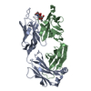
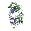
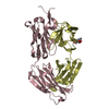
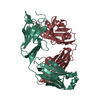
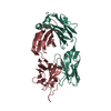
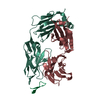
| Title | X-RAY CRYSTAL STRUCTURE OF RV2018 FROM MYCOBACTERIUM TUBERCULOSIS | ||||||
 Components Components | VAPBC49 | ||||||
 Keywords Keywords | DNA BINDING / MYCOBACTERIUM TUBERCULOSIS / TA SYSTEM | ||||||
| Function / homology | Putative antitoxin VapB45-like / : / Putative DNA-binding HTH domain / Protein of unknown function DUF433 / Protein of unknown function (DUF433) / Homeobox-like domain superfamily / Winged helix-like DNA-binding domain superfamily / Putative antitoxin VapB45 Function and homology information Function and homology information | ||||||
| Biological species |  MYCOBACTERIUM TUBERCULOSIS H37RV (bacteria) MYCOBACTERIUM TUBERCULOSIS H37RV (bacteria) | ||||||
| Method |  X-RAY DIFFRACTION / SYNCHROTRON / SIRAS / Resolution: 1.78 Å X-RAY DIFFRACTION / SYNCHROTRON / SIRAS / Resolution: 1.78 Å | ||||||
 Authors Authors | Holton, S.J. / Wilmanns, M. | ||||||
 Citation Citation | Journal: To be Published Title: Crystal Structure and DNA Binding Ability of Myco Tuberculosis Vapbc49 Anti-Toxin Protein Bacterium Authors: Holton, S.J. / Pogenberg, V. / Iborra, V. / Wilmanns, M. | ||||||
| History |
|
- Structure visualization
| Structure viewer | Molecule: MolmilJmol/JSmol |
|---|
- Downloads & links
-Download
| PDBx/mmCIF format | 5af3.cif.gz | 97.8 KB | Display | PDBx/mmCIF format |
|---|---|---|---|---|
| PDB format | pdb5af3.ent.gz | 75.8 KB | Display | PDB format |
| PDBx/mmJSON format | 5af3.json.gz | Tree view | PDBx/mmJSON format | |
| Others |  Other downloads Other downloads |
-Validation report
| Arichive directory | https://data.pdbj.org/pub/pdb/validation_reports/af/5af3ftp://data.pdbj.org/pub/pdb/validation_reports/af/5af3 | HTTPS FTP |
|---|
-Related structure data
| Similar structure data |
|---|


-Links
 PDBj
PDBj- Assembly
| Deposited unit | 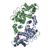
| ||||||||
|---|---|---|---|---|---|---|---|---|---|
| 1 |
| ||||||||
| Unit cell |
|
-Components
| #1: Protein | Mass: 26188.555 Da / Num. of mol.: 2 Source method: isolated from a genetically manipulated source Source: (gene. exp.) MYCOBACTERIUM TUBERCULOSIS H37RV (bacteria)Production host: #2: Chemical | ChemComp-GOL / |   Mass: 92.094 Da / Num. of mol.: 1 / Source method: obtained synthetically / Formula: C3H8O3 Mass: 92.094 Da / Num. of mol.: 1 / Source method: obtained synthetically / Formula: C3H8O3#3: Water | ChemComp-HOH / |  Mass: 18.015 Da / Num. of mol.: 289 / Source method: isolated from a natural source / Formula: H2O Mass: 18.015 Da / Num. of mol.: 289 / Source method: isolated from a natural source / Formula: H2O |
|---|
-Experimental details
-Experiment
| Experiment | Method: X-RAY DIFFRACTION / Number of used crystals: 1 |
|---|
- Sample preparation
| Crystal | Density Matthews: 2.89 Å3/Da / Density % sol: 57.13 % / Description: NONE |
|---|
-Data collection
| Diffraction | Mean temperature: 100 K |
|---|---|
| Diffraction source | Source: SYNCHROTRON / Site: BESSY  / Beamline: BW7A / Wavelength: 0.9792 / Beamline: BW7A / Wavelength: 0.9792 |
| Radiation | Protocol: SINGLE WAVELENGTH / Monochromatic (M) / Laue (L): M / Scattering type: x-ray |
| Radiation wavelength | Wavelength: 0.9792 Å / Relative weight: 1 |
| Reflection | Resolution: 1.77→40.96 Å / Num. obs: 53660 / % possible obs: 97.5 % / Observed criterion σ(I): 0 / Redundancy: 3.81 % / Rmerge(I) obs: 0.05 / Net I/σ(I): 10.73 |
| Reflection shell | Resolution: 1.77→1.87 Å / Redundancy: 3.55 % / Rmerge(I) obs: 0.46 / % possible all: 83.5 |
- Processing
| Software | Name: REFMAC / Version: 5.2.0005 / Classification: refinement | ||||||||||||||||||||
|---|---|---|---|---|---|---|---|---|---|---|---|---|---|---|---|---|---|---|---|---|---|
| Refinement | Method to determine structure: SIRAS Starting model: NONE Resolution: 1.78→70.89 Å / Cor.coef. Fo:Fc: 0.948 / Cor.coef. Fo:Fc free: 0.926 / SU B: 2.801 / SU ML: 0.089 / Cross valid method: THROUGHOUT / ESU R: 0.12 / ESU R Free: 0.123 / Stereochemistry target values: MAXIMUM LIKELIHOOD / Details: HYDROGENS HAVE BEEN ADDED IN THE RIDING POSITIONS.
| ||||||||||||||||||||
| Solvent computation | Ion probe radii: 0.8 Å / Shrinkage radii: 0.8 Å / VDW probe radii: 1.2 Å / Solvent model: MASK | ||||||||||||||||||||
| Displacement parameters | Biso mean: 26.25 Å2
| ||||||||||||||||||||
| Refinement step | Cycle: LAST / Resolution: 1.78→70.89 Å
|
