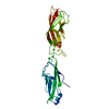
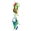
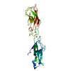
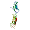
[English] 日本語
Yorodumi
- PDB-5abs: CRYSTAL STRUCTURE OF THE C-TERMINAL COILED-COIL DOMAIN OF CIN85 I... -

+
Open data


ID or keywords:

Loading...

-
Basic information

Entry
Database: PDB / ID: 5abs
TitleCRYSTAL STRUCTURE OF THE C-TERMINAL COILED-COIL DOMAIN OF CIN85 IN SPACE GROUP P321
ComponentsSH3 DOMAIN-CONTAINING KINASE-BINDING PROTEIN 1
KeywordsSIGNALING PROTEIN / CBL-INTERACTING PROTEIN OF 85 KDA / ADAPTER PROTEIN / COILED-COIL DOMAIN / B-CELL ANTIGEN RECEPTOR SIGNALING
Function / homology
Function and homology information


Reelin signalling pathway / positive regulation of B cell activation / endocytic vesicle / cytoskeleton organization / InlB-mediated entry of Listeria monocytogenes into host cell / Antigen activates B Cell Receptor (BCR) leading to generation of second messengers / actin filament organization / EGFR downregulation / cytoplasmic vesicle membrane / Negative regulation of MET activity ...Reelin signalling pathway / positive regulation of B cell activation / endocytic vesicle / cytoskeleton organization / InlB-mediated entry of Listeria monocytogenes into host cell / Antigen activates B Cell Receptor (BCR) leading to generation of second messengers / actin filament organization / EGFR downregulation / cytoplasmic vesicle membrane / Negative regulation of MET activity / SH3 domain binding / endocytosis / cell-cell junction / cell migration / Cargo recognition for clathrin-mediated endocytosis / cell-cell signaling / Clathrin-mediated endocytosis / regulation of cell shape / Potential therapeutics for SARS / cytoskeleton / neuron projection / focal adhesion / apoptotic process / ubiquitin protein ligase binding / synapse / plasma membrane / cytoplasm / cytosol
Similarity search - Function
SH3 domain-containing kinase-binding protein 1, first SH3 domain / SH3 domain-containing kinase-binding protein 1, second SH3 domain / SH3 domain-containing kinase-binding protein 1, third SH3 domain / Variant SH3 domain / Src homology 3 domains / SH3-like domain superfamily / Src homology 3 (SH3) domain profile. / SH3 domain
Similarity search - Domain/homology
SH3 domain-containing kinase-binding protein 1
Similarity search - Component
Biological speciesHOMO SAPIENS (human)
MethodX-RAY DIFFRACTION / SYNCHROTRON / SAD / Resolution: 1.74 Å
AuthorsWong, L. / Habeck, M. / Griesinger, C. / Becker, S.
CitationJournal: Sci.Signal. / Year: 2016
Title: The Adaptor Protein Cin85 Assembles Intracellular Signaling Clusters for B Cell Activation.
Authors: Kuhn, J. / Wong, L.E. / Pirkuliyeva, S. / Schulz, K. / Schwiegk, C. / Funfgeld, K.G. / Keppler, S. / Batista, F.D. / Urlaub, H. / Habeck, M. / Becker, S. / Griesinger, C. / Wienands, J.
History
DepositionAug 7, 2015Deposition site: PDBE / Processing site: PDBE
Revision 1.0Jul 13, 2016Provider: repository / Type: Initial release
Revision 1.1Jan 30, 2019Group: Data collection / Experimental preparation / Category: exptl_crystal_grow / Item: _exptl_crystal_grow.method
Revision 1.2Feb 6, 2019Group: Data collection / Experimental preparation / Category: exptl_crystal_grow / Item: _exptl_crystal_grow.temp

-
Structure visualization

Structure viewerMolecule:
MolmilJmol/JSmol

Downloads & links

-
Assembly

Deposited unit
A: SH3 DOMAIN-CONTAINING KINASE-BINDING PROTEIN 1
hetero molecules


Theoretical massNumber of molelcules
Total (without water)8,1475
Polymers7,8861
Non-polymers2624
Water25214
1
A: SH3 DOMAIN-CONTAINING KINASE-BINDING PROTEIN 1
hetero molecules

A: SH3 DOMAIN-CONTAINING KINASE-BINDING PROTEIN 1
hetero molecules

A: SH3 DOMAIN-CONTAINING KINASE-BINDING PROTEIN 1
hetero molecules


Theoretical massNumber of molelcules
Total (without water)24,44215
Polymers23,6573
Non-polymers78512
Water543
TypeNameSymmetry operationNumber
identity operation1_555x,y,z1
crystal symmetry operation3_665-x+y+1,-x+1,z1
crystal symmetry operation2_655-y+1,x-y,z1
Buried area7060 Å2
ΔGint-168.3 kcal/mol
Surface area12870 Å2
MethodPISA
Unit cell
Length a, b, c (Å)40.968, 40.968, 90.036
Angle α, β, γ (deg.)90.00, 90.00, 120.00
Int Tables number150
Space group name H-MP321
Components on special symmetry positions
IDModelComponents
11A-1003-

ZN

21A-1004-

ZN

-
Components

#1: Protein SH3 DOMAIN-CONTAINING KINASE-BINDING PROTEIN 1 / CD2-BINDING PROTEIN 3 / CD2BP3 / CBL-INTERACTING PROTEIN OF 85 KDA / HUMAN SRC FAMILY KINASE- ...CD2-BINDING PROTEIN 3 / CD2BP3 / CBL-INTERACTING PROTEIN OF 85 KDA / HUMAN SRC FAMILY KINASE-BINDING PROTEIN 1 / HSB-1


Mass: 7885.649 Da / Num. of mol.: 1 / Fragment: COILED-COIL DOMAIN, RESIDUES 599-662
Source method: isolated from a genetically manipulated source
Source: (gene. exp.) HOMO SAPIENS (human) / Production host: ESCHERICHIA COLI (E. coli) / Strain (production host): BL21(DE3) / References: UniProt: Q96B97
#2: Chemical
ChemComp-ZN / ZINC ION


Mass: 65.409 Da / Num. of mol.: 4 / Source method: obtained synthetically / Formula: Zn
#3: Water ChemComp-HOH / water


Mass: 18.015 Da / Num. of mol.: 14 / Source method: isolated from a natural source / Formula: H2O
Sequence detailsTHE N-TERMINAL GH IS DUE TO CLONING

-
Experimental details

-
Experiment

ExperimentMethod: X-RAY DIFFRACTION / Number of used crystals: 1

-
Sample preparation

CrystalDensity Matthews: 2.82 Å3/Da / Density % sol: 56.4 % / Description: NONE
Crystal growTemperature: 298 K / Method: vapor diffusion, sitting drop / pH: 7
Details: 0.05M ZN ACETATE, 20% PEG3350, VAPOR DIFFUSION, SITTING DROP, TEMPERATURE 298K, pH 7

-
Data collection

DiffractionMean temperature: 100 K
Diffraction sourceSource: SYNCHROTRON / Site: SLS / Beamline: X10SA / Wavelength: 1.214
DetectorType: DECTRIS PILATUS 6M / Detector: PIXEL / Date: Mar 7, 2015
RadiationProtocol: SINGLE WAVELENGTH / Monochromatic (M) / Laue (L): M / Scattering type: x-ray
Radiation wavelengthWavelength: 1.214 Å / Relative weight: 1
ReflectionResolution: 1.74→45.02 Å / Num. obs: 9541 / % possible obs: 99.6 % / Observed criterion σ(I): 2 / Redundancy: 17.9 % / Rmerge(I) obs: 0.1 / Net I/σ(I): 15.48
Reflection shellResolution: 1.74→1.83 Å / Redundancy: 16.88 % / Rmerge(I) obs: 0.92 / Mean I/σ(I) obs: 1.09 / % possible all: 97.6

-
Processing

Software
NameVersionClassification
REFMAC5.8.0124refinement
XDSdata reduction
SADABSdata scaling
SHELXCDEphasing
RefinementMethod to determine structure: SAD
Starting model: NONE

Resolution: 1.74→45.02 Å / Cor.coef. Fo:Fc: 0.953 / Cor.coef. Fo:Fc free: 0.918 / SU B: 3.897 / SU ML: 0.116 / Cross valid method: THROUGHOUT / ESU R: 0.13 / ESU R Free: 0.13 / Stereochemistry target values: MAXIMUM LIKELIHOOD / Details: HYDROGENS HAVE BEEN ADDED IN THE RIDING POSITIONS.
RfactorNum. reflection% reflectionSelection details
Rfree0.28327 993 10.4 %RANDOM
Rwork0.24397 ---
obs0.24796 8537 99.86 %-
Solvent computationIon probe radii: 0.8 Å / Shrinkage radii: 0.8 Å / VDW probe radii: 1.2 Å / Solvent model: MASK
Displacement parametersBiso mean: 43.917 Å2
Baniso -1Baniso -2Baniso -3
1-1.68 Å20.84 Å20 Å2
2--1.68 Å20 Å2
3----5.46 Å2
Refinement stepCycle: LAST / Resolution: 1.74→45.02 Å
ProteinNucleic acidLigandSolventTotal
Num. atoms520 0 4 14 538
Refine LS restraints
Refine-IDTypeDev idealDev ideal targetNumber
X-RAY DIFFRACTIONr_bond_refined_d0.0180.019539
X-RAY DIFFRACTIONr_bond_other_d0.0010.02549
X-RAY DIFFRACTIONr_angle_refined_deg1.9262.031723
X-RAY DIFFRACTIONr_angle_other_deg1.04831262
X-RAY DIFFRACTIONr_dihedral_angle_1_deg5.697569
X-RAY DIFFRACTIONr_dihedral_angle_2_deg32.19524.825
X-RAY DIFFRACTIONr_dihedral_angle_3_deg15.92415106
X-RAY DIFFRACTIONr_dihedral_angle_4_deg21.755156
X-RAY DIFFRACTIONr_chiral_restr0.1130.285
X-RAY DIFFRACTIONr_gen_planes_refined0.0070.02608
X-RAY DIFFRACTIONr_gen_planes_other0.0020.02108
X-RAY DIFFRACTIONr_nbd_refined
X-RAY DIFFRACTIONr_nbd_other
X-RAY DIFFRACTIONr_nbtor_refined
X-RAY DIFFRACTIONr_nbtor_other
X-RAY DIFFRACTIONr_xyhbond_nbd_refined
X-RAY DIFFRACTIONr_xyhbond_nbd_other
X-RAY DIFFRACTIONr_metal_ion_refined
X-RAY DIFFRACTIONr_metal_ion_other
X-RAY DIFFRACTIONr_symmetry_vdw_refined
X-RAY DIFFRACTIONr_symmetry_vdw_other
X-RAY DIFFRACTIONr_symmetry_hbond_refined
X-RAY DIFFRACTIONr_symmetry_hbond_other
X-RAY DIFFRACTIONr_symmetry_metal_ion_refined
X-RAY DIFFRACTIONr_symmetry_metal_ion_other
X-RAY DIFFRACTIONr_mcbond_it3.4473.909273
X-RAY DIFFRACTIONr_mcbond_other3.4463.9272
X-RAY DIFFRACTIONr_mcangle_it4.3685.818343
X-RAY DIFFRACTIONr_mcangle_other
X-RAY DIFFRACTIONr_scbond_it6.5584.772266
X-RAY DIFFRACTIONr_scbond_other
X-RAY DIFFRACTIONr_scangle_it
X-RAY DIFFRACTIONr_scangle_other
X-RAY DIFFRACTIONr_long_range_B_refined
X-RAY DIFFRACTIONr_long_range_B_other
X-RAY DIFFRACTIONr_rigid_bond_restr
X-RAY DIFFRACTIONr_sphericity_free
X-RAY DIFFRACTIONr_sphericity_bonded
LS refinement shellResolution: 1.738→1.83 Å / Total num. of bins used: 20
RfactorNum. reflection% reflection
Rfree0.364 76 -
Rwork0.472 603 -
obs--98.84 %

+
About Yorodumi

-
News

-
Feb 9, 2022. New format data for meta-information of EMDB entries

New format data for meta-information of EMDB entries

  • Version 3 of the EMDB header file is now the official format.
  • The previous official version 1.9 will be removed from the archive.

Related info.:EMDB header

External links:wwPDB to switch to version 3 of the EMDB data model

-
Aug 12, 2020. Covid-19 info

Covid-19 info

URL: https://pdbjlvh1.pdbj.org/emnavi/covid19.php

New page: Covid-19 featured information page in EM Navigator.

Related info.:Covid-19 info / Mar 5, 2020. Novel coronavirus structure data

+
Mar 5, 2020. Novel coronavirus structure data

Novel coronavirus structure data

Related info.:Yorodumi Speices / Aug 12, 2020. Covid-19 info

External links:COVID-19 featured content - PDBj / Molecule of the Month (242):Coronavirus Proteases

+
Jan 31, 2019. EMDB accession codes are about to change! (news from PDBe EMDB page)

EMDB accession codes are about to change! (news from PDBe EMDB page)

  • The allocation of 4 digits for EMDB accession codes will soon come to an end. Whilst these codes will remain in use, new EMDB accession codes will include an additional digit and will expand incrementally as the available range of codes is exhausted. The current 4-digit format prefixed with “EMD-” (i.e. EMD-XXXX) will advance to a 5-digit format (i.e. EMD-XXXXX), and so on. It is currently estimated that the 4-digit codes will be depleted around Spring 2019, at which point the 5-digit format will come into force.
  • The EM Navigator/Yorodumi systems omit the EMD- prefix.

Related info.:Q: What is EMD? / ID/Accession-code notation in Yorodumi/EM Navigator

External links:EMDB Accession Codes are Changing Soon! / Contact to PDBj

+
Jul 12, 2017. Major update of PDB

Major update of PDB

  • wwPDB released updated PDB data conforming to the new PDBx/mmCIF dictionary.
  • This is a major update changing the version number from 4 to 5, and with Remediation, in which all the entries are updated.
  • In this update, many items about electron microscopy experimental information are reorganized (e.g. em_software).
  • Now, EM Navigator and Yorodumi are based on the updated data.

External links:wwPDB Remediation / Enriched Model Files Conforming to OneDep Data Standards Now Available in the PDB FTP Archive

-
Yorodumi

Thousand views of thousand structures

  • Yorodumi is a browser for structure data from EMDB, PDB, SASBDB, etc.
  • This page is also the successor to EM Navigator detail page, and also detail information page/front-end page for Omokage search.
  • The word "yorodu" (or yorozu) is an old Japanese word meaning "ten thousand". "mi" (miru) is to see.

Related info.:EMDB / PDB / SASBDB / Comparison of 3 databanks / Yorodumi Search / Aug 31, 2016. New EM Navigator & Yorodumi / Yorodumi Papers / Jmol/JSmol / Function and homology information / Changes in new EM Navigator and Yorodumi

Read more