 Movie
Movie Controller

[English] 日本語
 Yorodumi
- PDB-5ab5: Crystal structure of Trypanosoma brucei SCP2-thiolase like protei... -
+ Open data

- Basic information
| Entry | Database: PDB / ID: 5ab5 | ||||||
|---|---|---|---|---|---|---|---|
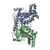
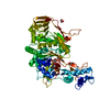
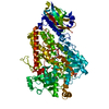
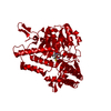
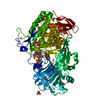
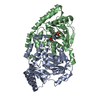
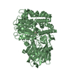
| Title | Crystal structure of Trypanosoma brucei SCP2-thiolase like protein (TbSLP) form-II. | ||||||
 Components Components | SCP2-THIOLASE LIKE PROTEIN | ||||||
 Keywords Keywords | TRANSPORT PROTEIN / COENZYME A / SCP2-THIOLASE / SCP2-THIOLASE-LIKE PROTEIN / MALONYL-COA DECARBOXYLASE / GENE KNOCKOUT / LIPID METABOLISM | ||||||
| Function / homology |  Function and homology information Function and homology informationacyltransferase activity, transferring groups other than amino-acyl groups Similarity search - Function | ||||||
| Biological species |  | ||||||
| Method |  X-RAY DIFFRACTION / SYNCHROTRON / MOLECULAR REPLACEMENT / Resolution: 2 Å X-RAY DIFFRACTION / SYNCHROTRON / MOLECULAR REPLACEMENT / Resolution: 2 Å | ||||||
 Authors Authors | Harijan, R.K. / Kiema, T.R. / Wierenga, R.K. | ||||||
 Citation Citation | Journal: Proteins / Year: 2016 Title: The Scp2-Thiolase-Like Protein (Slp) of Trypanosoma Brucei is an Enzyme Involved in Lipid Metabolism. Authors: Harijan, R.K. / Mazet, M. / Kiema, T.R. / Bouyssou, G. / Alexson, S.E.H. / Bergmann, U. / Moreau, P. / Michels, P.A.M. / Bringaud, F. / Wierenga, R.K. | ||||||
| History |
|
- Structure visualization
| Structure viewer | Molecule: MolmilJmol/JSmol |
|---|
- Downloads & links
-Download
| PDBx/mmCIF format | 5ab5.cif.gz | 162.8 KB | Display | PDBx/mmCIF format |
|---|---|---|---|---|
| PDB format | pdb5ab5.ent.gz | 130.6 KB | Display | PDB format |
| PDBx/mmJSON format | 5ab5.json.gz | Tree view | PDBx/mmJSON format | |
| Others |  Other downloads Other downloads |
-Validation report
| Arichive directory | https://data.pdbj.org/pub/pdb/validation_reports/ab/5ab5ftp://data.pdbj.org/pub/pdb/validation_reports/ab/5ab5 | HTTPS FTP |
|---|
-Related structure data


-Links
 PDBj
PDBj

- Assembly
| Deposited unit | 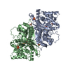
| ||||||||
|---|---|---|---|---|---|---|---|---|---|
| 1 |
| ||||||||
| Unit cell |
| ||||||||
| Noncrystallographic symmetry (NCS) | NCS oper: (Code: given Matrix: (-0.4979, -0.8672, -0.002531), Vector: |
-Components
| #1: Protein | Mass: 45365.898 Da / Num. of mol.: 2 Source method: isolated from a genetically manipulated source Source: (gene. exp.)  #2: Chemical | ChemComp-SO4 /   Mass: 96.063 Da / Num. of mol.: 13 / Source method: obtained synthetically / Formula: SO4 Mass: 96.063 Da / Num. of mol.: 13 / Source method: obtained synthetically / Formula: SO4#3: Water | ChemComp-HOH / |  Mass: 18.015 Da / Num. of mol.: 119 / Source method: isolated from a natural source / Formula: H2O Mass: 18.015 Da / Num. of mol.: 119 / Source method: isolated from a natural source / Formula: H2O |
|---|
-Experimental details
-Experiment
| Experiment | Method: X-RAY DIFFRACTION / Number of used crystals: 1 |
|---|
- Sample preparation
| Crystal | Density Matthews: 2.3 Å3/Da / Density % sol: 47.1 % / Description: NONE |
|---|---|
| Crystal grow | pH: 4.6 Details: 100 MM SODIUM ACETATE (PH 4.6), 2.0 M AMMONIUM SULPHATE |
-Data collection
| Diffraction | Mean temperature: 100 K |
|---|---|
| Diffraction source | Source: SYNCHROTRON / Site: Diamond  / Beamline: I03 / Wavelength: 0.97625 / Beamline: I03 / Wavelength: 0.97625 |
| Detector | Type: DECTRIS PILATUS 6M / Detector: PIXEL / Date: Oct 20, 2013 |
| Radiation | Monochromator: DOUBLE CRYSTAL / Protocol: SINGLE WAVELENGTH / Monochromatic (M) / Laue (L): M / Scattering type: x-ray |
| Radiation wavelength | Wavelength: 0.97625 Å / Relative weight: 1 |
| Reflection twin | Operator: H,-H-K,-L L / Fraction: 0.5 |
| Reflection | Resolution: 2→28.88 Å / Num. obs: 52628 / % possible obs: 99.8 % / Observed criterion σ(I): 1.3 / Redundancy: 5.1 % / Rmerge(I) obs: 0.1 / Net I/σ(I): 8.3 |
| Reflection shell | Resolution: 2→2.05 Å / Redundancy: 5.1 % / Rmerge(I) obs: 1.25 / Mean I/σ(I) obs: 1.3 / % possible all: 99 |
- Processing
| Software |
| |||||||||||||||||||||||||||||||||||||||||||||||||||||||||||||||||||||||||||||||||||||||||||||||||||||||||||||||||||||||||||||||||||||
|---|---|---|---|---|---|---|---|---|---|---|---|---|---|---|---|---|---|---|---|---|---|---|---|---|---|---|---|---|---|---|---|---|---|---|---|---|---|---|---|---|---|---|---|---|---|---|---|---|---|---|---|---|---|---|---|---|---|---|---|---|---|---|---|---|---|---|---|---|---|---|---|---|---|---|---|---|---|---|---|---|---|---|---|---|---|---|---|---|---|---|---|---|---|---|---|---|---|---|---|---|---|---|---|---|---|---|---|---|---|---|---|---|---|---|---|---|---|---|---|---|---|---|---|---|---|---|---|---|---|---|---|---|---|---|
| Refinement | Method to determine structure: MOLECULAR REPLACEMENT / Resolution: 2→28.877 Å / σ(F): 1.96 / Phase error: 30.58 / Stereochemistry target values: TWIN_LSQ_F
| |||||||||||||||||||||||||||||||||||||||||||||||||||||||||||||||||||||||||||||||||||||||||||||||||||||||||||||||||||||||||||||||||||||
| Solvent computation | Shrinkage radii: 0.9 Å / VDW probe radii: 1.11 Å / Solvent model: FLAT BULK SOLVENT MODEL | |||||||||||||||||||||||||||||||||||||||||||||||||||||||||||||||||||||||||||||||||||||||||||||||||||||||||||||||||||||||||||||||||||||
| Refinement step | Cycle: LAST / Resolution: 2→28.877 Å
| |||||||||||||||||||||||||||||||||||||||||||||||||||||||||||||||||||||||||||||||||||||||||||||||||||||||||||||||||||||||||||||||||||||
| Refine LS restraints |
| |||||||||||||||||||||||||||||||||||||||||||||||||||||||||||||||||||||||||||||||||||||||||||||||||||||||||||||||||||||||||||||||||||||
| LS refinement shell |
|
