 Movie
Movie Controller

[English] 日本語
 Yorodumi
- PDB-5aa4: Crystal structure of MltF from Pseudomonas aeruginosa in complex ... -
+ Open data

- Basic information
| Entry | Database: PDB / ID: 5aa4 | ||||||
|---|---|---|---|---|---|---|---|
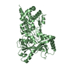
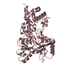
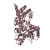
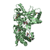
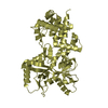
| Title | Crystal structure of MltF from Pseudomonas aeruginosa in complex with cell-wall tetrapeptide | ||||||
 Components Components | (MEMBRANE-BOUND LYTIC MUREIN TRANSGLYCOSYLASE F) x 2 | ||||||
 Keywords Keywords | LYASE / LYTIC TRANSGLYCOSILASE / CELL WALL RECYCLING | ||||||
| Function / homology |  Function and homology information Function and homology information: / peptidoglycan lytic transglycosylase activity / peptidoglycan catabolic process / cell outer membrane / cell wall organization / cell wall macromolecule catabolic process Similarity search - Function | ||||||
| Biological species |   PSEUDOMONAS AERUGINOSA (bacteria) PSEUDOMONAS AERUGINOSA (bacteria) | ||||||
| Method |  X-RAY DIFFRACTION / SYNCHROTRON / MOLECULAR REPLACEMENT / Resolution: 2.4 Å X-RAY DIFFRACTION / SYNCHROTRON / MOLECULAR REPLACEMENT / Resolution: 2.4 Å | ||||||
 Authors Authors | Dominguez-Gil, T. / Acebron, I. / Hermoso, J.A. | ||||||
 Citation Citation | Journal: Structure / Year: 2016 Title: Activation by Allostery in Cell-Wall Remodeling by a Modular Membrane-Bound Lytic Transglycosylase from Pseudomonas aeruginosa. Authors: Dominguez-Gil, T. / Lee, M. / Acebron-Avalos, I. / Mahasenan, K.V. / Hesek, D. / Dik, D.A. / Byun, B. / Lastochkin, E. / Fisher, J.F. / Mobashery, S. / Hermoso, J.A. | ||||||
| History |
|
- Structure visualization
| Structure viewer | Molecule: MolmilJmol/JSmol |
|---|
- Downloads & links
-Download
| PDBx/mmCIF format | 5aa4.cif.gz | 619.4 KB | Display | PDBx/mmCIF format |
|---|---|---|---|---|
| PDB format | pdb5aa4.ent.gz | 518.5 KB | Display | PDB format |
| PDBx/mmJSON format | 5aa4.json.gz | Tree view | PDBx/mmJSON format | |
| Others |  Other downloads Other downloads |
-Validation report
| Arichive directory | https://data.pdbj.org/pub/pdb/validation_reports/aa/5aa4ftp://data.pdbj.org/pub/pdb/validation_reports/aa/5aa4 | HTTPS FTP |
|---|
-Related structure data
| Related structure data | 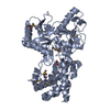 5a5xSC  5aa1C  5aa2C  5aa3C C: citing same article ( S: Starting model for refinement |
|---|---|
| Similar structure data |


-Links
 PDBj
PDBj- Assembly
| Deposited unit | 
| ||||||||
|---|---|---|---|---|---|---|---|---|---|
| 1 | 
| ||||||||
| 2 | 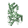
| ||||||||
| 3 | 
| ||||||||
| 4 | 
| ||||||||
| Unit cell |
|
-Components
| #1: Protein | Mass: 51113.395 Da / Num. of mol.: 2 Source method: isolated from a genetically manipulated source Source: (gene. exp.) PSEUDOMONAS AERUGINOSA (bacteria) / Strain: BWHPSA013 / Production host: References: UniProt: A0A077JMS0, UniProt: A0A1I9GEN8*PLUS, Lyases; Carbon-oxygen lyases; Acting on polysaccharides #2: Protein | Mass: 51143.418 Da / Num. of mol.: 2 Source method: isolated from a genetically manipulated source Source: (gene. exp.) PSEUDOMONAS AERUGINOSA (bacteria) / Strain: BWHPSA013 / Production host: References: UniProt: A0A077JMS0, UniProt: A0A1I9GEN9*PLUS, Lyases; Carbon-oxygen lyases; Acting on polysaccharides #3: Chemical | ChemComp-6X4 / [ |   Mass: 489.543 Da / Num. of mol.: 1 / Source method: obtained synthetically / Formula: C20H37N6O8 Mass: 489.543 Da / Num. of mol.: 1 / Source method: obtained synthetically / Formula: C20H37N6O8#4: Water | ChemComp-HOH / |  Mass: 18.015 Da / Num. of mol.: 160 / Source method: isolated from a natural source / Formula: H2O Mass: 18.015 Da / Num. of mol.: 160 / Source method: isolated from a natural source / Formula: H2OSequence details | FIRST NINE AMINOACIDS | |
|---|
-Experimental details
-Experiment
| Experiment | Method: X-RAY DIFFRACTION |
|---|
- Sample preparation
| Crystal | Density Matthews: 2.94 Å3/Da / Density % sol: 58.15 % / Description: NONE |
|---|
-Data collection
| Diffraction | Mean temperature: 100 K |
|---|---|
| Diffraction source | Source: SYNCHROTRON / Site: ALBA  / Beamline: XALOC / Wavelength: 1 / Beamline: XALOC / Wavelength: 1 |
| Detector | Type: DECTRIS PILATUS 6M / Detector: PIXEL / Details: ELLIPTICALLY BENT MIRROR |
| Radiation | Monochromator: SI(111) CHANNEL-CUT, CRYOCOOLED / Protocol: SINGLE WAVELENGTH / Monochromatic (M) / Laue (L): M / Scattering type: x-ray |
| Radiation wavelength | Wavelength: 1 Å / Relative weight: 1 |
| Reflection | Resolution: 2.4→48.72 Å / Num. obs: 94338 / % possible obs: 98.5 % / Observed criterion σ(I): 0 / Redundancy: 6.7 % / Rmerge(I) obs: 0.09 / Net I/σ(I): 13.8 |
| Reflection shell | Resolution: 2.4→2.44 Å / Redundancy: 6.7 % / Rmerge(I) obs: 1.04 / Mean I/σ(I) obs: 1.7 / % possible all: 99.9 |
- Processing
| Software |
| |||||||||||||||||||||||||||||||||||||||||||||||||||||||||||||||||||||||||||||||||||||||||||||||||||||||||||||||||||||||||||||||||||||||||||||||||||||||||||||||||||||||||||||||||||||||||||||||||||||||||||||||||||||||||
|---|---|---|---|---|---|---|---|---|---|---|---|---|---|---|---|---|---|---|---|---|---|---|---|---|---|---|---|---|---|---|---|---|---|---|---|---|---|---|---|---|---|---|---|---|---|---|---|---|---|---|---|---|---|---|---|---|---|---|---|---|---|---|---|---|---|---|---|---|---|---|---|---|---|---|---|---|---|---|---|---|---|---|---|---|---|---|---|---|---|---|---|---|---|---|---|---|---|---|---|---|---|---|---|---|---|---|---|---|---|---|---|---|---|---|---|---|---|---|---|---|---|---|---|---|---|---|---|---|---|---|---|---|---|---|---|---|---|---|---|---|---|---|---|---|---|---|---|---|---|---|---|---|---|---|---|---|---|---|---|---|---|---|---|---|---|---|---|---|---|---|---|---|---|---|---|---|---|---|---|---|---|---|---|---|---|---|---|---|---|---|---|---|---|---|---|---|---|---|---|---|---|---|---|---|---|---|---|---|---|---|---|---|---|---|---|---|---|---|
| Refinement | Method to determine structure: MOLECULAR REPLACEMENT Starting model: PDB ENTRY 5A5X Resolution: 2.4→48.569 Å / SU ML: 0.35 / σ(F): 1.33 / Phase error: 26.73 / Stereochemistry target values: ML
| |||||||||||||||||||||||||||||||||||||||||||||||||||||||||||||||||||||||||||||||||||||||||||||||||||||||||||||||||||||||||||||||||||||||||||||||||||||||||||||||||||||||||||||||||||||||||||||||||||||||||||||||||||||||||
| Solvent computation | Shrinkage radii: 0.9 Å / VDW probe radii: 1.11 Å / Solvent model: FLAT BULK SOLVENT MODEL | |||||||||||||||||||||||||||||||||||||||||||||||||||||||||||||||||||||||||||||||||||||||||||||||||||||||||||||||||||||||||||||||||||||||||||||||||||||||||||||||||||||||||||||||||||||||||||||||||||||||||||||||||||||||||
| Refinement step | Cycle: LAST / Resolution: 2.4→48.569 Å
| |||||||||||||||||||||||||||||||||||||||||||||||||||||||||||||||||||||||||||||||||||||||||||||||||||||||||||||||||||||||||||||||||||||||||||||||||||||||||||||||||||||||||||||||||||||||||||||||||||||||||||||||||||||||||
| Refine LS restraints |
| |||||||||||||||||||||||||||||||||||||||||||||||||||||||||||||||||||||||||||||||||||||||||||||||||||||||||||||||||||||||||||||||||||||||||||||||||||||||||||||||||||||||||||||||||||||||||||||||||||||||||||||||||||||||||
| LS refinement shell |
|
