 ムービー
ムービー コントローラー


+ データを開く

- 基本情報
基本情報
| 登録情報 | データベース: PDB / ID: 5a8f | ||||||
|---|---|---|---|---|---|---|---|
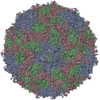
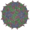
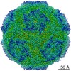
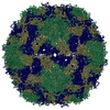
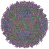
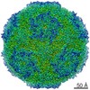
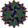
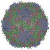
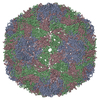
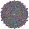
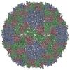
| タイトル | Structure and genome release mechanism of human cardiovirus Saffold virus-3 | ||||||
 要素 要素 |
| ||||||
 キーワード キーワード | VIRAL PROTEIN / SAFFOLD / VIRUS / CARDIOVIRUS / PICORNAVIRALES / A / ALTERED / VIRION / PARTICLE / CAPSID / GENOME / RNA / SSRNA | ||||||
| 機能・相同性 |  機能・相同性情報 機能・相同性情報host cell nucleolus / symbiont-mediated suppression of host mRNA export from nucleus / picornain 3C / T=pseudo3 icosahedral viral capsid / host cell cytoplasmic vesicle membrane / channel activity / monoatomic ion transmembrane transport / RNA helicase activity / RNA helicase / RNA-directed RNA polymerase ...host cell nucleolus / symbiont-mediated suppression of host mRNA export from nucleus / picornain 3C / T=pseudo3 icosahedral viral capsid / host cell cytoplasmic vesicle membrane / channel activity / monoatomic ion transmembrane transport / RNA helicase activity / RNA helicase / RNA-directed RNA polymerase / cysteine-type endopeptidase activity / viral RNA genome replication / RNA-directed RNA polymerase activity / symbiont entry into host cell / DNA-templated transcription / virion attachment to host cell / structural molecule activity / ATP hydrolysis activity / proteolysis / RNA binding / zinc ion binding / ATP binding 類似検索 - 分子機能 | ||||||
| 生物種 |  SAFFOLD VIRUS (ウイルス) SAFFOLD VIRUS (ウイルス) | ||||||
| 手法 | 電子顕微鏡法 / 単粒子再構成法 / クライオ電子顕微鏡法 / 解像度: 10.6 Å | ||||||
 データ登録者 データ登録者 | Mullapudi, E. / Novacek, J. / Palkova, L. / Kulich, P. / Lindberg, M. / vanKuppeveld, F.J.M. / Plevka, P. | ||||||
 引用 引用 | ジャーナル: J Virol / 年: 2016 タイトル: Structure and Genome Release Mechanism of the Human Cardiovirus Saffold Virus 3. 著者: Edukondalu Mullapudi / Jiří Nováček / Lenka Pálková / Pavel Kulich / A Michael Lindberg / Frank J M van Kuppeveld / Pavel Plevka /    要旨: In order to initiate an infection, viruses need to deliver their genomes into cells. This involves uncoating the genome and transporting it to the cytoplasm. The process of genome delivery is not ...In order to initiate an infection, viruses need to deliver their genomes into cells. This involves uncoating the genome and transporting it to the cytoplasm. The process of genome delivery is not well understood for nonenveloped viruses. We address this gap in our current knowledge by studying the uncoating of the nonenveloped human cardiovirus Saffold virus 3 (SAFV-3) of the family Picornaviridae SAFVs cause diseases ranging from gastrointestinal disorders to meningitis. We present a structure of a native SAFV-3 virion determined to 2.5 Å by X-ray crystallography and an 11-Å-resolution cryo-electron microscopy reconstruction of an "altered" particle that is primed for genome release. The altered particles are expanded relative to the native virus and contain pores in the capsid that might serve as channels for the release of VP4 subunits, N termini of VP1, and the RNA genome. Unlike in the related enteroviruses, pores in SAFV-3 are located roughly between the icosahedral 3- and 5-fold axes at an interface formed by two VP1 and one VP3 subunit. Furthermore, in native conditions many cardioviruses contain a disulfide bond formed by cysteines that are separated by just one residue. The disulfide bond is located in a surface loop of VP3. We determined the structure of the SAFV-3 virion in which the disulfide bonds are reduced. Disruption of the bond had minimal effect on the structure of the loop, but it increased the stability and decreased the infectivity of the virus. Therefore, compounds specifically disrupting or binding to the disulfide bond might limit SAFV infection. IMPORTANCE: A capsid assembled from viral proteins protects the virus genome during transmission from one cell to another. However, when a virus enters a cell the virus genome has to be released from ...IMPORTANCE: A capsid assembled from viral proteins protects the virus genome during transmission from one cell to another. However, when a virus enters a cell the virus genome has to be released from the capsid in order to initiate infection. This process is not well understood for nonenveloped viruses. We address this gap in our current knowledge by studying the genome release of Human Saffold virus 3 Saffold viruses cause diseases ranging from gastrointestinal disorders to meningitis. We show that before the genome is released, the Saffold virus 3 particle expands, and holes form in the previously compact capsid. These holes serve as channels for the release of the genome and small capsid proteins VP4 that in related enteroviruses facilitate subsequent transport of the virus genome into the cell cytoplasm. | ||||||
| 履歴 |
|
- 構造の表示
構造の表示
| ムービー |
ムービービューア |
|---|---|
| 構造ビューア | 分子: MolmilJmol/JSmol |
- ダウンロードとリンク
-ダウンロード
| PDBx/mmCIF形式 | 5a8f.cif.gz | 141.8 KB | 表示 | PDBx/mmCIF形式 |
|---|---|---|---|---|
| PDB形式 | pdb5a8f.ent.gz | 109.7 KB | 表示 | PDB形式 |
| PDBx/mmJSON形式 | 5a8f.json.gz | ツリー表示 | PDBx/mmJSON形式 | |
| その他 |  その他のダウンロード その他のダウンロード |
-検証レポート
| アーカイブディレクトリ | https://data.pdbj.org/pub/pdb/validation_reports/a8/5a8fftp://data.pdbj.org/pub/pdb/validation_reports/a8/5a8f | HTTPS FTP |
|---|
-関連構造データ


-リンク
 PDBj
PDBj

- 集合体
集合体
| 登録構造単位 | 
|
|---|---|
| 1 | x 60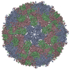
|
| 2 |
|
| 3 | x 5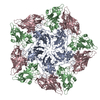
|
| 4 | x 6
|
| 5 | 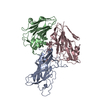
|
| 対称性 | 点対称性: (シェーンフリース記号: I (正20面体型対称)) |
-要素
| #1: タンパク質 | 分子量: 24785.914 Da / 分子数: 1 / 由来タイプ: 天然 / 由来: (天然) SAFFOLD VIRUS (ウイルス) / 参照: UniProt: C0MHL9 |
|---|---|
| #2: タンパク質 | 分子量: 20893.826 Da / 分子数: 1 / 由来タイプ: 天然 / 由来: (天然) SAFFOLD VIRUS (ウイルス) / 参照: UniProt: E3TMG9 |
| #3: タンパク質 | 分子量: 28698.160 Da / 分子数: 1 / 由来タイプ: 天然 / 由来: (天然) SAFFOLD VIRUS (ウイルス) / 参照: UniProt: C0MHL9 |
| Has protein modification | Y |
-実験情報
-実験
| 実験 | 手法: 電子顕微鏡法 |
|---|---|
| EM実験 | 試料の集合状態: PARTICLE / 3次元再構成法: 単粒子再構成法 |
- 試料調製
試料調製
| 構成要素 | 名称: A PARTICLE OF SAFFOLD VIRUS-3 / タイプ: VIRUS |
|---|---|
| 緩衝液 | 名称: 20MM HEPES, 150MM NACL / pH: 7.5 / 詳細: 20MM HEPES, 150MM NACL |
| 試料 | 濃度: 2 mg/ml / 包埋: NO / シャドウイング: NO / 染色: NO / 凍結: YES |
| 試料支持 | 詳細: HOLEY CARBON |
| 急速凍結 | 装置: FEI VITROBOT MARK IV / 凍結剤: ETHANE 詳細: CRYOGEN - LIQUID ETHANE, VITROBOT MARK IV, BLOT FOR 2 SECONDS |
- 電子顕微鏡撮影
| 実験機器 |  モデル: Tecnai F20 / 画像提供: FEI Company |
|---|---|
| 顕微鏡 | モデル: FEI TECNAI F20 / 日付: 2014年9月3日 |
| 電子銃 | 電子線源:  FIELD EMISSION GUN / 加速電圧: 200 kV / 照射モード: SPOT SCAN FIELD EMISSION GUN / 加速電圧: 200 kV / 照射モード: SPOT SCAN |
| 電子レンズ | モード: BRIGHT FIELD / 倍率(公称値): 55000 X / 最大 デフォーカス(公称値): 3950 nm / 最小 デフォーカス(公称値): 2330 nm / Cs: 2 mm |
| 試料ホルダ | 傾斜角・最大: 0 ° |
| 撮影 | 電子線照射量: 20 e/Å2 / フィルム・検出器のモデル: FEI EAGLE (4k x 4k) |
| 画像スキャン | デジタル画像の数: 264 |
- 解析
解析
| EMソフトウェア |
| ||||||||||||||||||||
|---|---|---|---|---|---|---|---|---|---|---|---|---|---|---|---|---|---|---|---|---|---|
| CTF補正 | 詳細: EACH PARTICLE | ||||||||||||||||||||
| 対称性 | 点対称性: I (正20面体型対称) | ||||||||||||||||||||
| 3次元再構成 | 手法: PROJECTION MATCHING, FOURIER METHODS / 解像度: 10.6 Å / 粒子像の数: 14028 / ピクセルサイズ(公称値): 2.2176 Å / ピクセルサイズ(実測値): 2.2176 Å 詳細: SUBMISSION BASED ON EXPERIMENTAL DATA FROM EMDB EMD-3097. (DEPOSITION ID: 13604). 対称性のタイプ: POINT | ||||||||||||||||||||
| 原子モデル構築 | プロトコル: OTHER / 空間: REAL / 詳細: METHOD--LOCAL CORRELATION | ||||||||||||||||||||
| 精密化 | 最高解像度: 10.6 Å | ||||||||||||||||||||
| 精密化ステップ | サイクル: LAST / 最高解像度: 10.6 Å
|
