 Movie
Movie Controller

[English] 日本語
 Yorodumi
- PDB-5a52: The crystal structure of Arabidopsis thaliana CAR1 in complex wit... -
+ Open data

- Basic information
| Entry | Database: PDB / ID: 5a52 | ||||||
|---|---|---|---|---|---|---|---|
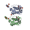
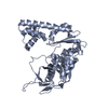
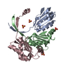
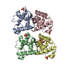
| Title | The crystal structure of Arabidopsis thaliana CAR1 in complex with one calcium ion | ||||||
 Components Components | CALCIUM-DEPENDENT LIPID-BINDING DOMAIN-CONTAINING PROTEIN | ||||||
 Keywords Keywords | LIPID BINDING PROTEIN / CALCIUM BINDING PROTEIN / C2 DOMAIN | ||||||
| Function / homology |  Function and homology information Function and homology informationpositive regulation of abscisic acid-activated signaling pathway / abscisic acid-activated signaling pathway / GTPase activator activity / phospholipid binding / calcium ion binding / lipid binding / protein homodimerization activity / nucleus / plasma membrane Similarity search - Function | ||||||
| Biological species |  | ||||||
| Method |  X-RAY DIFFRACTION / SYNCHROTRON / MOLECULAR REPLACEMENT / Resolution: 1.65 Å X-RAY DIFFRACTION / SYNCHROTRON / MOLECULAR REPLACEMENT / Resolution: 1.65 Å | ||||||
 Authors Authors | Fernandez, D. / Marquez, J.A. | ||||||
 Citation Citation | Journal: Proc.Natl.Acad.Sci.USA / Year: 2016 Title: Calcium-Dependent Oligomerization of Car Proteins at Cell Membrane Modulates Aba Signaling. Authors: Diaz, M. / Sanchez-Barrena, M.J. / Gonzalez-Rubio, J.M. / Rodriguez, L. / Fernandez, D. / Antoni, R. / Yunta, C. / Belda-Palazon, B. / Gonzalez-Guzman, M. / Peirats-Llobet, M. / Menendez, M. ...Authors: Diaz, M. / Sanchez-Barrena, M.J. / Gonzalez-Rubio, J.M. / Rodriguez, L. / Fernandez, D. / Antoni, R. / Yunta, C. / Belda-Palazon, B. / Gonzalez-Guzman, M. / Peirats-Llobet, M. / Menendez, M. / Boskovic, J. / Marquez, J.A. / Rodriguez, P.L. / Albert, A. | ||||||
| History |
|
- Structure visualization
| Structure viewer | Molecule: MolmilJmol/JSmol |
|---|
- Downloads & links
-Download
| PDBx/mmCIF format | 5a52.cif.gz | 46.6 KB | Display | PDBx/mmCIF format |
|---|---|---|---|---|
| PDB format | pdb5a52.ent.gz | 32.8 KB | Display | PDB format |
| PDBx/mmJSON format | 5a52.json.gz | Tree view | PDBx/mmJSON format | |
| Others |  Other downloads Other downloads |
-Validation report
| Arichive directory | https://data.pdbj.org/pub/pdb/validation_reports/a5/5a52ftp://data.pdbj.org/pub/pdb/validation_reports/a5/5a52 | HTTPS FTP |
|---|
-Related structure data
| Related structure data |  5a4xC 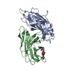 5a50C  5a51C  4v29S C: citing same article ( S: Starting model for refinement |
|---|---|
| Similar structure data |


-Links
 PDBj
PDBj


- Assembly
| Deposited unit | 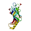
| ||||||||
|---|---|---|---|---|---|---|---|---|---|
| 1 | 
| ||||||||
| Unit cell |
|
-Components
| #1: Protein | Mass: 18867.793 Da / Num. of mol.: 1 Source method: isolated from a genetically manipulated source Source: (gene. exp.)  | ||||||
|---|---|---|---|---|---|---|---|
| #2: Chemical |   Mass: 96.063 Da / Num. of mol.: 2 / Source method: obtained synthetically / Formula: SO4 Mass: 96.063 Da / Num. of mol.: 2 / Source method: obtained synthetically / Formula: SO4#3: Chemical | ChemComp-GOL / |   Mass: 92.094 Da / Num. of mol.: 1 / Source method: obtained synthetically / Formula: C3H8O3 Mass: 92.094 Da / Num. of mol.: 1 / Source method: obtained synthetically / Formula: C3H8O3#4: Chemical | ChemComp-CA / |   Mass: 40.078 Da / Num. of mol.: 1 / Source method: obtained synthetically / Formula: Ca Mass: 40.078 Da / Num. of mol.: 1 / Source method: obtained synthetically / Formula: Ca#5: Water | ChemComp-HOH / |  Mass: 18.015 Da / Num. of mol.: 96 / Source method: isolated from a natural source / Formula: H2O Mass: 18.015 Da / Num. of mol.: 96 / Source method: isolated from a natural source / Formula: H2O |
-Experimental details
-Experiment
| Experiment | Method: X-RAY DIFFRACTION / Number of used crystals: 1 |
|---|
- Sample preparation
| Crystal | Density Matthews: 2.38 Å3/Da / Density % sol: 48.26 % / Description: NONE |
|---|---|
| Crystal grow | Details: 0.5 M MGSO4, 0.5 M HEPES PH 7.0 AND 1.6 M LITHIUM SULFATE |
-Data collection
| Diffraction | Mean temperature: 100 K |
|---|---|
| Diffraction source | Source: SYNCHROTRON / Site: ESRF  / Beamline: ID23-1 / Wavelength: 0.98 / Beamline: ID23-1 / Wavelength: 0.98 |
| Detector | Type: DECTRIS PIXEL / Detector: PIXEL |
| Radiation | Protocol: SINGLE WAVELENGTH / Monochromatic (M) / Laue (L): M / Scattering type: x-ray |
| Radiation wavelength | Wavelength: 0.98 Å / Relative weight: 1 |
| Reflection | Resolution: 1.65→23.23 Å / Num. obs: 22034 / % possible obs: 99.4 % / Observed criterion σ(I): 0 / Redundancy: 5.9 % / Biso Wilson estimate: 21.35 Å2 / Rmerge(I) obs: 0.06 / Net I/σ(I): 20.79 |
| Reflection shell | Resolution: 1.65→1.75 Å / Redundancy: 5.9 % / Rmerge(I) obs: 0.7 / Mean I/σ(I) obs: 3.1 / % possible all: 99.1 |
- Processing
| Software |
| |||||||||||||||||||||||||||||||||||||||||||||||||||||||||||||||
|---|---|---|---|---|---|---|---|---|---|---|---|---|---|---|---|---|---|---|---|---|---|---|---|---|---|---|---|---|---|---|---|---|---|---|---|---|---|---|---|---|---|---|---|---|---|---|---|---|---|---|---|---|---|---|---|---|---|---|---|---|---|---|---|---|
| Refinement | Method to determine structure: MOLECULAR REPLACEMENT Starting model: PDB ENTRY 4V29 Resolution: 1.65→23.231 Å / SU ML: 0.16 / σ(F): 1.36 / Phase error: 23.94 / Stereochemistry target values: ML
| |||||||||||||||||||||||||||||||||||||||||||||||||||||||||||||||
| Solvent computation | Shrinkage radii: 0.9 Å / VDW probe radii: 1.11 Å / Solvent model: FLAT BULK SOLVENT MODEL | |||||||||||||||||||||||||||||||||||||||||||||||||||||||||||||||
| Refinement step | Cycle: LAST / Resolution: 1.65→23.231 Å
| |||||||||||||||||||||||||||||||||||||||||||||||||||||||||||||||
| Refine LS restraints |
| |||||||||||||||||||||||||||||||||||||||||||||||||||||||||||||||
| LS refinement shell |
|
