 Movie
Movie Controller

[English] 日本語
 Yorodumi
- PDB-5a2b: Crystal Structure of Anoxybacillus Alpha-amylase Provides Insight... -
+ Open data

- Basic information
| Entry | Database: PDB / ID: 5a2b | |||||||||
|---|---|---|---|---|---|---|---|---|---|---|
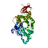
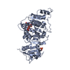
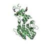
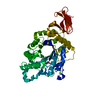
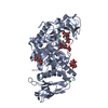
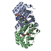
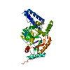
| Title | Crystal Structure of Anoxybacillus Alpha-amylase Provides Insights into a New Glycosyl Hydrolase Subclass | |||||||||
 Components Components | ANOXYBACILLUS ALPHA-AMYLASE | |||||||||
 Keywords Keywords | HYDROLASE / CALCIUM-BINDING SITE / GEOBACILLUS / GLYCOSYL HYDROLASE | |||||||||
| Function / homology |  Function and homology information Function and homology informationalpha-amylase / alpha-amylase activity / carbohydrate metabolic process / calcium ion binding Similarity search - Function | |||||||||
| Biological species |  ANOXYBACILLUS SP. (bacteria) ANOXYBACILLUS SP. (bacteria) | |||||||||
| Method |  X-RAY DIFFRACTION / MOLECULAR REPLACEMENT / Resolution: 1.85 Å X-RAY DIFFRACTION / MOLECULAR REPLACEMENT / Resolution: 1.85 Å | |||||||||
 Authors Authors | Ng, C.L. / Chai, K.P. / Othman, N.F. / Teh, A.H. / Ho, K.L. / Chan, K.G. / Goh, K.M. | |||||||||
 Citation Citation | Journal: Sci.Rep. / Year: 2016 Title: Crystal Structure of Anoxybacillus Alpha-Amylase Provides Insights Into Maltose Binding of a New Glycosyl Hydrolase Subclass. Authors: Chai, K.P. / Othman, N.F.B. / Teh, A. / Ho, K.L. / Chan, K. / Shamsir, M.S. / Goh, K.M. / Ng, C.L. | |||||||||
| History |
| |||||||||
| Remark 700 | SHEET DETERMINATION METHOD: DSSP THE SHEETS PRESENTED AS "AA" IN EACH CHAIN ON SHEET RECORDS BELOW ... SHEET DETERMINATION METHOD: DSSP THE SHEETS PRESENTED AS "AA" IN EACH CHAIN ON SHEET RECORDS BELOW IS ACTUALLY AN 8-STRANDED BARREL THIS IS REPRESENTED BY A 9-STRANDED SHEET IN WHICH THE FIRST AND LAST STRANDS ARE IDENTICAL. |
- Structure visualization
| Structure viewer | Molecule: MolmilJmol/JSmol |
|---|
- Downloads & links
-Download
| PDBx/mmCIF format | 5a2b.cif.gz | 220.2 KB | Display | PDBx/mmCIF format |
|---|---|---|---|---|
| PDB format | pdb5a2b.ent.gz | 175.3 KB | Display | PDB format |
| PDBx/mmJSON format | 5a2b.json.gz | Tree view | PDBx/mmJSON format | |
| Others |  Other downloads Other downloads |
-Validation report
| Arichive directory | https://data.pdbj.org/pub/pdb/validation_reports/a2/5a2bftp://data.pdbj.org/pub/pdb/validation_reports/a2/5a2b | HTTPS FTP |
|---|
-Related structure data
| Related structure data |  5a2aC  5a2cC 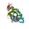 4e2oS C: citing same article ( S: Starting model for refinement |
|---|---|
| Similar structure data |


-Links
 PDBj
PDBj


- Assembly
| Deposited unit | 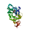
| ||||||||
|---|---|---|---|---|---|---|---|---|---|
| 1 |
| ||||||||
| Unit cell |
|
-Components
| #1: Protein | Mass: 58164.398 Da / Num. of mol.: 1 / Fragment: TRUNCATED PROTEIN, RESIDUES 24-478 Source method: isolated from a genetically manipulated source Details: CATALYTIC DOMAIN A WITH TIM BARREL FOLD (RESIDUES 26 TO 139,187 TO 393), DOMAIN B (RESIDUES 140 TO 186), AND DOMAIN C WITH AN ALL-ALPHA-BETA FOLD (RESIDUES 394 TO 475) Source: (gene. exp.) ANOXYBACILLUS SP. (bacteria) / Strain: SK3-4 / Description: ISOLATED FROM SUNGAI KLAH HOT SPRING MALAYSIA / Plasmid: PET28A / Production host: | ||
|---|---|---|---|
| #2: Polysaccharide | alpha-D-glucopyranose-(1-4)-alpha-D-glucopyranose / alpha-maltose  Source method: isolated from a genetically manipulated source Details: oligosaccharide / References: alpha-maltose | ||
| #3: Chemical | ChemComp-CA /   Mass: 40.078 Da / Num. of mol.: 4 / Source method: obtained synthetically / Formula: Ca Mass: 40.078 Da / Num. of mol.: 4 / Source method: obtained synthetically / Formula: Ca#4: Water | ChemComp-HOH / |  Mass: 18.015 Da / Num. of mol.: 412 / Source method: isolated from a natural source / Formula: H2O Mass: 18.015 Da / Num. of mol.: 412 / Source method: isolated from a natural source / Formula: H2O |
-Experimental details
-Experiment
| Experiment | Method: X-RAY DIFFRACTION / Number of used crystals: 1 |
|---|
- Sample preparation
| Crystal | Density Matthews: 2.48 Å3/Da / Density % sol: 50.5 % / Description: NONE |
|---|---|
| Crystal grow | pH: 6.5 Details: 0.2 M CALCIUM ACETATE 0.1 M SODIUM CACODYLATE (PH 6.5) 18% (W/V) POLYETHYLENE GLYCOL 8, 000 20 MM MALTOSE |
-Data collection
| Diffraction | Mean temperature: 100 K |
|---|---|
| Diffraction source | Source: ROTATING ANODE / Type: RIGAKU MICROMAX-007 HF / Wavelength: 1.5148 |
| Detector | Type: RIGAKU R-AXIS IV / Detector: IMAGE PLATE / Date: Jul 10, 2014 |
| Radiation | Protocol: SINGLE WAVELENGTH / Monochromatic (M) / Laue (L): M / Scattering type: x-ray |
| Radiation wavelength | Wavelength: 1.5148 Å / Relative weight: 1 |
| Reflection | Resolution: 1.85→19.6 Å / Num. obs: 43446 / % possible obs: 94.5 % / Observed criterion σ(I): 1.8 / Redundancy: 3.8 % / Rmerge(I) obs: 0.06 / Net I/σ(I): 15.7 |
| Reflection shell | Resolution: 1.85→1.89 Å / Redundancy: 2.8 % / Rmerge(I) obs: 0.52 / Mean I/σ(I) obs: 1.8 / % possible all: 83.6 |
- Processing
| Software |
| ||||||||||||||||||||||||||||||||||||||||||||||||||||||||||||||||||||||||||||||||||||||||||||||||||||||||||||||||||||||||||||||||||||||||||||||||||||||||||||||||||||||||||||||||||||||
|---|---|---|---|---|---|---|---|---|---|---|---|---|---|---|---|---|---|---|---|---|---|---|---|---|---|---|---|---|---|---|---|---|---|---|---|---|---|---|---|---|---|---|---|---|---|---|---|---|---|---|---|---|---|---|---|---|---|---|---|---|---|---|---|---|---|---|---|---|---|---|---|---|---|---|---|---|---|---|---|---|---|---|---|---|---|---|---|---|---|---|---|---|---|---|---|---|---|---|---|---|---|---|---|---|---|---|---|---|---|---|---|---|---|---|---|---|---|---|---|---|---|---|---|---|---|---|---|---|---|---|---|---|---|---|---|---|---|---|---|---|---|---|---|---|---|---|---|---|---|---|---|---|---|---|---|---|---|---|---|---|---|---|---|---|---|---|---|---|---|---|---|---|---|---|---|---|---|---|---|---|---|---|---|
| Refinement | Method to determine structure: MOLECULAR REPLACEMENT Starting model: 4E2O Resolution: 1.85→63.35 Å / Cor.coef. Fo:Fc: 0.969 / Cor.coef. Fo:Fc free: 0.951 / SU B: 7.101 / SU ML: 0.092 / Cross valid method: THROUGHOUT / ESU R: 0.124 / ESU R Free: 0.118 / Stereochemistry target values: MAXIMUM LIKELIHOOD Details: HYDROGENS HAVE BEEN ADDED IN THE RIDING POSITIONS. HYDROGENS HAVE BEEN ADDED IN THE RIDING POSITIONS U VALUES WITH TLS ADDED
| ||||||||||||||||||||||||||||||||||||||||||||||||||||||||||||||||||||||||||||||||||||||||||||||||||||||||||||||||||||||||||||||||||||||||||||||||||||||||||||||||||||||||||||||||||||||
| Solvent computation | Ion probe radii: 0.8 Å / Shrinkage radii: 0.8 Å / VDW probe radii: 1.2 Å / Solvent model: MASK | ||||||||||||||||||||||||||||||||||||||||||||||||||||||||||||||||||||||||||||||||||||||||||||||||||||||||||||||||||||||||||||||||||||||||||||||||||||||||||||||||||||||||||||||||||||||
| Displacement parameters | Biso mean: 25.357 Å2
| ||||||||||||||||||||||||||||||||||||||||||||||||||||||||||||||||||||||||||||||||||||||||||||||||||||||||||||||||||||||||||||||||||||||||||||||||||||||||||||||||||||||||||||||||||||||
| Refinement step | Cycle: LAST / Resolution: 1.85→63.35 Å
| ||||||||||||||||||||||||||||||||||||||||||||||||||||||||||||||||||||||||||||||||||||||||||||||||||||||||||||||||||||||||||||||||||||||||||||||||||||||||||||||||||||||||||||||||||||||
| Refine LS restraints |
|