 Movie
Movie Controller

[English] 日本語
 Yorodumi
- PDB-3lx6: Crystal structure of putative dna cytosine methylase from shigell... -
+ Open data

- Basic information
| Entry | Database: PDB / ID: 3lx6 | ||||||
|---|---|---|---|---|---|---|---|
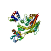
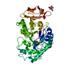
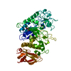
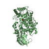
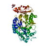
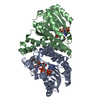
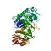
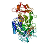
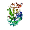
| Title | Crystal structure of putative dna cytosine methylase from shigella flexneri 2a str. 2457T | ||||||
 Components Components | Cytosine-specific methyltransferase | ||||||
 Keywords Keywords | TRANSFERASE / structural genomics / dcm / Restriction system / PSI-2 / Protein Structure Initiative / New York SGX Research Center for Structural Genomics / NYSGXRC | ||||||
| Function / homology | DNA Methylase; Chain A, domain 2 - #30 / DNA Methylase; Chain A, domain 2 / Vaccinia Virus protein VP39 / Alpha-Beta Complex / Rossmann fold / 3-Layer(aba) Sandwich / Alpha Beta / :  Function and homology information Function and homology information | ||||||
| Biological species |  Shigella flexneri 2a (bacteria) Shigella flexneri 2a (bacteria) | ||||||
| Method |  X-RAY DIFFRACTION / SYNCHROTRON / SAD / Resolution: 2.29 Å X-RAY DIFFRACTION / SYNCHROTRON / SAD / Resolution: 2.29 Å | ||||||
 Authors Authors | Ramagopal, U.A. / Toro, R. / Burley, S.K. / Almo, S.C. / New York SGX Research Center for Structural Genomics (NYSGXRC) | ||||||
 Citation Citation | Journal: To be Published Title: Crystal structure of putative dna cytosine methylase from shigella flexneri 2a str. 2457T Authors: Ramagopal, U.A. / Toro, R. / Burley, S.K. / Almo, S.C. | ||||||
| History |
|
- Structure visualization
| Structure viewer | Molecule: MolmilJmol/JSmol |
|---|
- Downloads & links
-Download
| PDBx/mmCIF format | 3lx6.cif.gz | 155.2 KB | Display | PDBx/mmCIF format |
|---|---|---|---|---|
| PDB format | pdb3lx6.ent.gz | 120.9 KB | Display | PDB format |
| PDBx/mmJSON format | 3lx6.json.gz | Tree view | PDBx/mmJSON format | |
| Others |  Other downloads Other downloads |
-Validation report
| Arichive directory | https://data.pdbj.org/pub/pdb/validation_reports/lx/3lx6ftp://data.pdbj.org/pub/pdb/validation_reports/lx/3lx6 | HTTPS FTP |
|---|
-Related structure data


-Links
 PDBj
PDBj- Assembly
| Deposited unit | 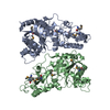
| ||||||||
|---|---|---|---|---|---|---|---|---|---|
| 1 | 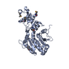
| ||||||||
| 2 | 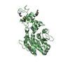
| ||||||||
| Unit cell |
| ||||||||
| Details | unknown |
-Components
| #1: Protein | Mass: 47123.414 Da / Num. of mol.: 2 Source method: isolated from a genetically manipulated source Source: (gene. exp.) Shigella flexneri 2a (bacteria) / Strain: 2457T / Gene: dcm, S2100, SF2005, SF2457T_1495 / Plasmid: BC-pSGX4(BC) / Production host: References: UniProt: E3Y0J2, DNA (cytosine-5-)-methyltransferase #2: Water | ChemComp-HOH / |  Mass: 18.015 Da / Num. of mol.: 29 / Source method: isolated from a natural source / Formula: H2O Mass: 18.015 Da / Num. of mol.: 29 / Source method: isolated from a natural source / Formula: H2OHas protein modification | Y | |
|---|
-Experimental details
-Experiment
| Experiment | Method: X-RAY DIFFRACTION / Number of used crystals: 1 |
|---|
- Sample preparation
| Crystal | Density Matthews: 2.72 Å3/Da / Density % sol: 54.78 % |
|---|---|
| Crystal grow | Temperature: 298 K / Method: vapor diffusion, sitting drop / pH: 7.5 Details: 0.1 M Hepes pH 7.5, 10% PEG 8K, 8% Ethylene Glycol, Vapor diffusion, Sitting drop, temperature 298K |
-Data collection
| Diffraction | Mean temperature: 100 K | |||||||||||||||||||||||||||||||||||||||||||||||||||||||||||||||||||||||||||||||||||||||||||||||||||||||||||||||||||||||||||||||||||||||||||||||||||
|---|---|---|---|---|---|---|---|---|---|---|---|---|---|---|---|---|---|---|---|---|---|---|---|---|---|---|---|---|---|---|---|---|---|---|---|---|---|---|---|---|---|---|---|---|---|---|---|---|---|---|---|---|---|---|---|---|---|---|---|---|---|---|---|---|---|---|---|---|---|---|---|---|---|---|---|---|---|---|---|---|---|---|---|---|---|---|---|---|---|---|---|---|---|---|---|---|---|---|---|---|---|---|---|---|---|---|---|---|---|---|---|---|---|---|---|---|---|---|---|---|---|---|---|---|---|---|---|---|---|---|---|---|---|---|---|---|---|---|---|---|---|---|---|---|---|---|---|---|
| Diffraction source | Source: SYNCHROTRON / Site: APS  / Beamline: 24-ID-E / Wavelength: 0.9793 Å / Beamline: 24-ID-E / Wavelength: 0.9793 Å | |||||||||||||||||||||||||||||||||||||||||||||||||||||||||||||||||||||||||||||||||||||||||||||||||||||||||||||||||||||||||||||||||||||||||||||||||||
| Detector | Type: ADSC QUANTUM 315 / Detector: CCD / Date: Nov 5, 2009 | |||||||||||||||||||||||||||||||||||||||||||||||||||||||||||||||||||||||||||||||||||||||||||||||||||||||||||||||||||||||||||||||||||||||||||||||||||
| Radiation | Protocol: SINGLE WAVELENGTH / Scattering type: x-ray | |||||||||||||||||||||||||||||||||||||||||||||||||||||||||||||||||||||||||||||||||||||||||||||||||||||||||||||||||||||||||||||||||||||||||||||||||||
| Radiation wavelength | Wavelength: 0.9793 Å / Relative weight: 1 | |||||||||||||||||||||||||||||||||||||||||||||||||||||||||||||||||||||||||||||||||||||||||||||||||||||||||||||||||||||||||||||||||||||||||||||||||||
| Reflection | Resolution: 2.29→50 Å / Num. obs: 43909 / % possible obs: 96.8 % / Redundancy: 3.2 % / Rmerge(I) obs: 0.083 / Χ2: 1.366 / Net I/σ(I): 12.4 | |||||||||||||||||||||||||||||||||||||||||||||||||||||||||||||||||||||||||||||||||||||||||||||||||||||||||||||||||||||||||||||||||||||||||||||||||||
| Reflection shell |
|
- Processing
| Software |
| |||||||||||||||||||||||||||||||||||||||||||||||||||||||||||||||||||||||||||
|---|---|---|---|---|---|---|---|---|---|---|---|---|---|---|---|---|---|---|---|---|---|---|---|---|---|---|---|---|---|---|---|---|---|---|---|---|---|---|---|---|---|---|---|---|---|---|---|---|---|---|---|---|---|---|---|---|---|---|---|---|---|---|---|---|---|---|---|---|---|---|---|---|---|---|---|---|
| Refinement | Method to determine structure: SAD / Resolution: 2.29→31.26 Å / Cor.coef. Fo:Fc: 0.926 / Cor.coef. Fo:Fc free: 0.898 / WRfactor Rfree: 0.302 / WRfactor Rwork: 0.257 / Occupancy max: 1 / Occupancy min: 0.5 / FOM work R set: 0.763 / SU B: 18.944 / SU ML: 0.211 / SU R Cruickshank DPI: 0.323 / SU Rfree: 0.254 / TLS residual ADP flag: LIKELY RESIDUAL / Cross valid method: THROUGHOUT / σ(F): 0 / ESU R: 0.323 / ESU R Free: 0.254 / Stereochemistry target values: MAXIMUM LIKELIHOOD Details: HYDROGENS HAVE BEEN ADDED IN THE RIDING POSITIONS U VALUES : RESIDUAL ONLY
| |||||||||||||||||||||||||||||||||||||||||||||||||||||||||||||||||||||||||||
| Solvent computation | Ion probe radii: 0.8 Å / Shrinkage radii: 0.8 Å / VDW probe radii: 1.4 Å / Solvent model: BABINET MODEL WITH MASK | |||||||||||||||||||||||||||||||||||||||||||||||||||||||||||||||||||||||||||
| Displacement parameters | Biso max: 83.47 Å2 / Biso mean: 32.744 Å2 / Biso min: 13.02 Å2
| |||||||||||||||||||||||||||||||||||||||||||||||||||||||||||||||||||||||||||
| Refinement step | Cycle: LAST / Resolution: 2.29→31.26 Å
| |||||||||||||||||||||||||||||||||||||||||||||||||||||||||||||||||||||||||||
| Refine LS restraints |
| |||||||||||||||||||||||||||||||||||||||||||||||||||||||||||||||||||||||||||
| LS refinement shell | Resolution: 2.292→2.351 Å / Total num. of bins used: 20
| |||||||||||||||||||||||||||||||||||||||||||||||||||||||||||||||||||||||||||
| Refinement TLS params. | Method: refined / Refine-ID: X-RAY DIFFRACTION
| |||||||||||||||||||||||||||||||||||||||||||||||||||||||||||||||||||||||||||
| Refinement TLS group |
|
