 Movie
Movie Controller


+ Open data

- Basic information
| Entry | Database: PDB / ID: 5a23 | ||||||
|---|---|---|---|---|---|---|---|
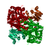
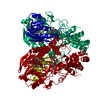
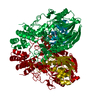
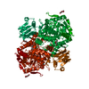
| Title | SdsA sulfatase triclinic form | ||||||
 Components Components | SDS HYDROLASE SDSA1 | ||||||
 Keywords Keywords | HYDROLASE / SDSA SULFATASE / POLYMORPHS | ||||||
| Function / homology |  Function and homology information Function and homology informationlinear primary-alkylsulfatase / linear primary-alkylsulfatase activity / dodecyl sulfate metabolic process / outer membrane-bounded periplasmic space / protein dimerization activity / metal ion binding / identical protein binding Similarity search - Function | ||||||
| Biological species |   PSEUDOMONAS AERUGINOSA (bacteria) PSEUDOMONAS AERUGINOSA (bacteria) | ||||||
| Method |  X-RAY DIFFRACTION / SYNCHROTRON / MOLECULAR REPLACEMENT / Resolution: 2.41 Å X-RAY DIFFRACTION / SYNCHROTRON / MOLECULAR REPLACEMENT / Resolution: 2.41 Å | ||||||
 Authors Authors | De la Mora, E. / Flores-Hernandez, E. / Jakoncik, J. / Stojanoff, V. / Sanchez-Puig, N. / Moreno, A. | ||||||
 Citation Citation | Journal: J.Appl.Crystallogr. / Year: 2015 Title: Sdsa Polymorph Isolation and Improvement of Their Crystal Quality Using Nonconventional Crystallization Techniques Authors: De La Mora, E. / Flores-Hernandez, E. / Jakoncic, J. / Stojanoff, V. / Siliqi, D. / Sanchez-Puig, N. / Moreno, A. | ||||||
| History |
|
- Structure visualization
| Structure viewer | Molecule: MolmilJmol/JSmol |
|---|
- Downloads & links
-Download
| PDBx/mmCIF format | 5a23.cif.gz | 889.8 KB | Display | PDBx/mmCIF format |
|---|---|---|---|---|
| PDB format | pdb5a23.ent.gz | 744.5 KB | Display | PDB format |
| PDBx/mmJSON format | 5a23.json.gz | Tree view | PDBx/mmJSON format | |
| Others |  Other downloads Other downloads |
-Validation report
| Arichive directory | https://data.pdbj.org/pub/pdb/validation_reports/a2/5a23ftp://data.pdbj.org/pub/pdb/validation_reports/a2/5a23 | HTTPS FTP |
|---|
-Related structure data
| Related structure data |  5aijC  5ajlC  2cg3S S: Starting model for refinement C: citing same article ( |
|---|---|
| Similar structure data |


-Links
 PDBj
PDBj


- Assembly
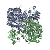
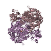
| Deposited unit | 
| ||||||||||||||||
|---|---|---|---|---|---|---|---|---|---|---|---|---|---|---|---|---|---|
| 1 | 
| ||||||||||||||||
| 2 | 
| ||||||||||||||||
| Unit cell |
| ||||||||||||||||
| Noncrystallographic symmetry (NCS) | NCS oper:
|
-Components
| #1: Protein | Mass: 72673.031 Da / Num. of mol.: 4 Source method: isolated from a genetically manipulated source Source: (gene. exp.) PSEUDOMONAS AERUGINOSA (bacteria) / Production host: #2: Chemical | ChemComp-ZN /   Mass: 65.409 Da / Num. of mol.: 8 / Source method: obtained synthetically / Formula: Zn Mass: 65.409 Da / Num. of mol.: 8 / Source method: obtained synthetically / Formula: Zn |
|---|
-Experimental details
-Experiment
| Experiment | Method: X-RAY DIFFRACTION / Number of used crystals: 1 |
|---|
- Sample preparation
| Crystal | Density Matthews: 3.32 Å3/Da / Density % sol: 63.1 % / Description: NONE |
|---|---|
| Crystal grow | pH: 7 Details: 10% PEG 4000, 10% ISOPROPANOL, 100MM LICL, 100 MM SODIUM CITRATE PH 6. |
-Data collection
| Diffraction | Mean temperature: 100 K |
|---|---|
| Diffraction source | Source: SYNCHROTRON / Site: APS  / Beamline: 19-BM / Wavelength: 0.9791 / Beamline: 19-BM / Wavelength: 0.9791 |
| Detector | Type: ADSC CCD / Detector: CCD / Date: Mar 13, 2014 |
| Radiation | Protocol: SINGLE WAVELENGTH / Monochromatic (M) / Laue (L): M / Scattering type: x-ray |
| Radiation wavelength | Wavelength: 0.9791 Å / Relative weight: 1 |
| Reflection | Resolution: 2.41→50 Å / Num. obs: 95678 / % possible obs: 71.8 % / Observed criterion σ(I): 1.65 / Redundancy: 2.97 % / Rmerge(I) obs: 0.06 / Net I/σ(I): 15.34 |
| Reflection shell | Resolution: 2.41→2.46 Å / Redundancy: 1.94 % / Rmerge(I) obs: 0.33 / Mean I/σ(I) obs: 1.65 / % possible all: 59.7 |
- Processing
| Software |
| ||||||||||||||||||||||||||||||||||||||||||||||||||||||||||||||||||||||||||||||||||||||||||||||||||||||||||||||||||||||||||||||||||||||||||||||||||||||||||||||||||||||||||||||||||||||
|---|---|---|---|---|---|---|---|---|---|---|---|---|---|---|---|---|---|---|---|---|---|---|---|---|---|---|---|---|---|---|---|---|---|---|---|---|---|---|---|---|---|---|---|---|---|---|---|---|---|---|---|---|---|---|---|---|---|---|---|---|---|---|---|---|---|---|---|---|---|---|---|---|---|---|---|---|---|---|---|---|---|---|---|---|---|---|---|---|---|---|---|---|---|---|---|---|---|---|---|---|---|---|---|---|---|---|---|---|---|---|---|---|---|---|---|---|---|---|---|---|---|---|---|---|---|---|---|---|---|---|---|---|---|---|---|---|---|---|---|---|---|---|---|---|---|---|---|---|---|---|---|---|---|---|---|---|---|---|---|---|---|---|---|---|---|---|---|---|---|---|---|---|---|---|---|---|---|---|---|---|---|---|---|
| Refinement | Method to determine structure: MOLECULAR REPLACEMENT Starting model: PDB ENTRY 2CG3 Resolution: 2.41→118.77 Å / Cor.coef. Fo:Fc: 0.901 / Cor.coef. Fo:Fc free: 0.853 / SU B: 27.741 / SU ML: 0.297 / Cross valid method: THROUGHOUT / ESU R: 0.884 / ESU R Free: 0.392 / Stereochemistry target values: MAXIMUM LIKELIHOOD / Details: HYDROGENS HAVE BEEN ADDED IN THE RIDING POSITIONS
| ||||||||||||||||||||||||||||||||||||||||||||||||||||||||||||||||||||||||||||||||||||||||||||||||||||||||||||||||||||||||||||||||||||||||||||||||||||||||||||||||||||||||||||||||||||||
| Solvent computation | Ion probe radii: 0.8 Å / Shrinkage radii: 0.8 Å / VDW probe radii: 1.2 Å / Solvent model: MASK | ||||||||||||||||||||||||||||||||||||||||||||||||||||||||||||||||||||||||||||||||||||||||||||||||||||||||||||||||||||||||||||||||||||||||||||||||||||||||||||||||||||||||||||||||||||||
| Displacement parameters | Biso mean: 46.865 Å2
| ||||||||||||||||||||||||||||||||||||||||||||||||||||||||||||||||||||||||||||||||||||||||||||||||||||||||||||||||||||||||||||||||||||||||||||||||||||||||||||||||||||||||||||||||||||||
| Refinement step | Cycle: LAST / Resolution: 2.41→118.77 Å
| ||||||||||||||||||||||||||||||||||||||||||||||||||||||||||||||||||||||||||||||||||||||||||||||||||||||||||||||||||||||||||||||||||||||||||||||||||||||||||||||||||||||||||||||||||||||
| Refine LS restraints |
|