[English] 日本語
 Yorodumi
- PDB-5a22: Structure of the L protein of vesicular stomatitis virus from ele... -
+ Open data
- Basic information
| Entry | Database: PDB / ID: 5a22 | ||||||
|---|---|---|---|---|---|---|---|
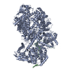
| Title | Structure of the L protein of vesicular stomatitis virus from electron cryomicroscopy | ||||||
 Components Components | VESICULAR STOMATITIS VIRUS L POLYMERASE | ||||||
 Keywords Keywords | TRANSFERASE / RNA-DEPENDENT RNA POLYMERASE / RNA CAPPING / CRYOEM SINGLE- PARTICLE ANALYSIS | ||||||
| Function / homology |  Function and homology information Function and homology informationNNS virus cap methyltransferase / GDP polyribonucleotidyltransferase / negative stranded viral RNA replication / viral transcription / Hydrolases; Acting on acid anhydrides; In phosphorus-containing anhydrides / virion component / mRNA 5'-cap (guanine-N7-)-methyltransferase activity / host cell cytoplasm / RNA-directed RNA polymerase / RNA-directed RNA polymerase activity ...NNS virus cap methyltransferase / GDP polyribonucleotidyltransferase / negative stranded viral RNA replication / viral transcription / Hydrolases; Acting on acid anhydrides; In phosphorus-containing anhydrides / virion component / mRNA 5'-cap (guanine-N7-)-methyltransferase activity / host cell cytoplasm / RNA-directed RNA polymerase / RNA-directed RNA polymerase activity / GTPase activity / ATP binding / metal ion binding Similarity search - Function | ||||||
| Biological species |  VESICULAR STOMATITIS VIRUS VESICULAR STOMATITIS VIRUS | ||||||
| Method | ELECTRON MICROSCOPY / single particle reconstruction / cryo EM / Resolution: 3.8 Å | ||||||
 Authors Authors | Liang, B. / Li, Z. / Jenni, S. / Rameh, A.A. / Morin, B.M. / Grant, T. / Grigorieff, N. / Harrison, S.C. / Whelan, S.P.J. | ||||||
 Citation Citation |  Journal: Cell / Year: 2015 Journal: Cell / Year: 2015Title: Structure of the L Protein of Vesicular Stomatitis Virus from Electron Cryomicroscopy. Authors: Bo Liang / Zongli Li / Simon Jenni / Amal A Rahmeh / Benjamin M Morin / Timothy Grant / Nikolaus Grigorieff / Stephen C Harrison / Sean P J Whelan /  Abstract: The large (L) proteins of non-segmented, negative-strand RNA viruses, a group that includes Ebola and rabies viruses, catalyze RNA-dependent RNA polymerization with viral ribonucleoprotein as ...The large (L) proteins of non-segmented, negative-strand RNA viruses, a group that includes Ebola and rabies viruses, catalyze RNA-dependent RNA polymerization with viral ribonucleoprotein as template, a non-canonical sequence of capping and methylation reactions, and polyadenylation of viral messages. We have determined by electron cryomicroscopy the structure of the vesicular stomatitis virus (VSV) L protein. The density map, at a resolution of 3.8 Å, has led to an atomic model for nearly all of the 2109-residue polypeptide chain, which comprises three enzymatic domains (RNA-dependent RNA polymerase [RdRp], polyribonucleotidyl transferase [PRNTase], and methyltransferase) and two structural domains. The RdRp resembles the corresponding enzymatic regions of dsRNA virus polymerases and influenza virus polymerase. A loop from the PRNTase (capping) domain projects into the catalytic site of the RdRp, where it appears to have the role of a priming loop and to couple product elongation to large-scale conformational changes in L. | ||||||
| History |
|
- Structure visualization
| Movie |
 Movie viewer Movie viewer |
|---|---|
| Structure viewer | Molecule:  Molmil Molmil Jmol/JSmol Jmol/JSmol |
- Downloads & links
- Download
| PDBx/mmCIF format |  5a22.cif.gz 5a22.cif.gz | 761.5 KB | Display |  PDBx/mmCIF format PDBx/mmCIF format |
|---|---|---|---|---|
| PDB format |  pdb5a22.ent.gz pdb5a22.ent.gz | 631.3 KB | Display |  PDB format PDB format |
| PDBx/mmJSON format |  5a22.json.gz 5a22.json.gz | Tree view |  PDBx/mmJSON format PDBx/mmJSON format | |
| Others |  Other downloads Other downloads |
-Validation report
| Arichive directory |  https://data.pdbj.org/pub/pdb/validation_reports/a2/5a22 https://data.pdbj.org/pub/pdb/validation_reports/a2/5a22 ftp://data.pdbj.org/pub/pdb/validation_reports/a2/5a22 ftp://data.pdbj.org/pub/pdb/validation_reports/a2/5a22 | HTTPS FTP |
|---|
-Related structure data
| Related structure data |  6337MC M: map data used to model this data C: citing same article ( |
|---|---|
| Similar structure data |
- Links
Links
- Assembly
| Deposited unit | 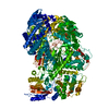
|
|---|---|
| 1 |
|
- Components
| #1: Protein | Mass: 241313.859 Da / Num. of mol.: 1 Source method: isolated from a genetically manipulated source Source: (gene. exp.)  VESICULAR STOMATITIS VIRUS / Strain: INDIANA / Variant: SAN JUAN ISOLATE / Plasmid: PFASTBAC DUAL (INVITROGEN) / Cell line (production host): Sf21 / Production host: VESICULAR STOMATITIS VIRUS / Strain: INDIANA / Variant: SAN JUAN ISOLATE / Plasmid: PFASTBAC DUAL (INVITROGEN) / Cell line (production host): Sf21 / Production host:  | ||
|---|---|---|---|
| #2: Chemical | | Has protein modification | Y | |
-Experimental details
-Experiment
| Experiment | Method: ELECTRON MICROSCOPY |
|---|---|
| EM experiment | Aggregation state: PARTICLE / 3D reconstruction method: single particle reconstruction |
- Sample preparation
| Component | Name: VESICULAR STOMATITIS VIRUS RNA- DEPENDENT RNA POLYMERASE (L) BOUND TO PHOSPHOPROTEIN P FRAGMENT Type: COMPLEX |
|---|---|
| Buffer solution | Name: 25 MM HEPES, 250 MM NACL, 6 MM MGSO4, 0.5 MM TCEP / pH: 7.4 / Details: 25 MM HEPES, 250 MM NACL, 6 MM MGSO4, 0.5 MM TCEP |
| Specimen | Conc.: 0.35 mg/ml / Embedding applied: NO / Shadowing applied: NO / Staining applied: NO / Vitrification applied: YES |
| Specimen support | Details: HOLEY CARBON |
| Vitrification | Instrument: FEI VITROBOT MARK I / Cryogen name: ETHANE / Details: LIQUID ETHANE |
- Electron microscopy imaging
| Experimental equipment |  Model: Tecnai F20 / Image courtesy: FEI Company |
|---|---|
| Microscopy | Model: FEI TECNAI F20 / Date: Aug 1, 2014 / Details: GOOD MICROGRAPHS WERE SELECTED FOR DIGITISATION |
| Electron gun | Electron source:  FIELD EMISSION GUN / Accelerating voltage: 200 kV / Illumination mode: OTHER FIELD EMISSION GUN / Accelerating voltage: 200 kV / Illumination mode: OTHER |
| Electron lens | Mode: OTHER / Nominal magnification: 29000 X / Calibrated magnification: 40410 X / Nominal defocus max: 2300 nm / Nominal defocus min: 900 nm / Cs: 2 mm |
| Specimen holder | Temperature: 100 K |
| Image recording | Electron dose: 100 e/Å2 / Film or detector model: GATAN K2 (4k x 4k) |
| Image scans | Num. digital images: 1272 |
| Radiation wavelength | Relative weight: 1 |
- Processing
| EM software |
| ||||||||||||||||
|---|---|---|---|---|---|---|---|---|---|---|---|---|---|---|---|---|---|
| CTF correction | Details: INDIVIDUAL PARTICLES | ||||||||||||||||
| Symmetry | Point symmetry: C1 (asymmetric) | ||||||||||||||||
| 3D reconstruction | Method: CROSS-COMMON LINES / Resolution: 3.8 Å / Num. of particles: 74940 / Nominal pixel size: 1.72 Å / Actual pixel size: 1.24 Å Details: FREALIGN WAS USED FOR REFINEMENT AND THREE- DIMENSIONAL CLASSIFICATION. SEGMENTS A1159-A1171, A1210-A1226, A1308-1334, A1387-A1395, A1512-A1516, A1534-A1541 ARE IN POOR DENSITY, AND THE ...Details: FREALIGN WAS USED FOR REFINEMENT AND THREE- DIMENSIONAL CLASSIFICATION. SEGMENTS A1159-A1171, A1210-A1226, A1308-1334, A1387-A1395, A1512-A1516, A1534-A1541 ARE IN POOR DENSITY, AND THE CHAIN TRACE IN THOSE REGIONS IS APPROXIMATE. STEREOCHEMISTRY OF ZN A3000, A3001 COORDINATION IS POOR. DEPOSITED STRUCTURE FACTORS USED FOR REFINEMENT OF THIS ENTRY ARE FROM SOLVENT FLATTENED EM MAP EMD CODE 6337 AFTER PLACING THE DENSITY INTO A CELL WITH DIMESIONS 112.000, 143.000, 106.000 AND ANGLES 90.00,90.00,90.00. APPLY THE FOLLOWING TRANSFORMATION MATRIX TO THE COORDINATES IN ORDER TO PLACE THE MODEL INTO THE DENSITY CALCULATED FROM THE DEPOSITED STRUCTURE FACTORS: MODEL2SF 1 -0.492787 0.222962 -0.841098 139.886 MODEL2SF 1 -0.456450 -0.889187 0.031721 169.772 MODEL2SF 1 -0.740818 0.399552 0.539950 33.800 Symmetry type: POINT | ||||||||||||||||
| Atomic model building | Protocol: FLEXIBLE FIT / Space: RECIPROCAL / Target criteria: Maximum likelihood / Details: METHOD--PHENIX.REFINE | ||||||||||||||||
| Refinement | Highest resolution: 3.8 Å | ||||||||||||||||
| Refinement step | Cycle: LAST / Highest resolution: 3.8 Å
|
 Movie
Movie Controller




 PDBj
PDBj


