 Movie
Movie Controller

[English] 日本語
 Yorodumi
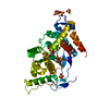
- PDB-4zx2: Co-crystal structures of PP5 in complex with 5-methyl-7-oxabicycl... -
+ Open data

- Basic information
| Entry | Database: PDB / ID: 4zx2 | ||||||||||||
|---|---|---|---|---|---|---|---|---|---|---|---|---|---|
| Title | Co-crystal structures of PP5 in complex with 5-methyl-7-oxabicyclo[2.2.1]heptane-2,3-dicarboxylic acid | ||||||||||||
 Components Components | Serine/threonine-protein phosphatase 5 | ||||||||||||
 Keywords Keywords | Hydrolase/Hydrolase Inhibitor / Protein phosphatase 5 / Hydrolase-Hydrolase Inhibitor complex | ||||||||||||
| Function / homology |  Function and homology information Function and homology informationpeptidyl-serine dephosphorylation / response to arachidonate / positive regulation of nuclear receptor-mediated glucocorticoid signaling pathway / proximal dendrite / mitogen-activated protein kinase kinase kinase binding / protein-serine/threonine phosphatase / response to morphine / cellular response to cadmium ion / protein serine/threonine phosphatase activity / phosphatase activity ...peptidyl-serine dephosphorylation / response to arachidonate / positive regulation of nuclear receptor-mediated glucocorticoid signaling pathway / proximal dendrite / mitogen-activated protein kinase kinase kinase binding / protein-serine/threonine phosphatase / response to morphine / cellular response to cadmium ion / protein serine/threonine phosphatase activity / phosphatase activity / protein folding chaperone complex / G-protein alpha-subunit binding / protein serine/threonine kinase inhibitor activity / negative regulation of MAPK cascade / phosphoprotein phosphatase activity / ESR-mediated signaling / Hsp70 protein binding / Hsp90 protein binding / ADP binding / response to lead ion / tau protein binding / cellular response to hydrogen peroxide / Negative regulation of MAPK pathway / MAPK cascade / double-strand break repair / mitotic cell cycle / Recruitment and ATM-mediated phosphorylation of repair and signaling proteins at DNA double strand breaks / microtubule binding / perikaryon / positive regulation of canonical NF-kappaB signal transduction / lipid binding / negative regulation of apoptotic process / DNA-templated transcription / protein-containing complex binding / protein-containing complex / RNA binding / nucleoplasm / ATP binding / metal ion binding / identical protein binding / nucleus / plasma membrane / cytosol Similarity search - Function | ||||||||||||
| Biological species |  Homo sapiens (human) Homo sapiens (human) | ||||||||||||
| Method |  X-RAY DIFFRACTION / SYNCHROTRON / MOLECULAR REPLACEMENT / Resolution: 1.23 Å X-RAY DIFFRACTION / SYNCHROTRON / MOLECULAR REPLACEMENT / Resolution: 1.23 Å | ||||||||||||
 Authors Authors | Chattopadhyay, D. / Swingle, M.R. / Salter, E.A. / Wierzbicki, A. / Honkanen, R.E. | ||||||||||||
| Funding support |  United States, 3items United States, 3items
| ||||||||||||
 Citation Citation | Journal: Biochem. Pharmacol. / Year: 2016 Title: Crystal structures and mutagenesis of PPP-family ser/thr protein phosphatases elucidate the selectivity of cantharidin and novel norcantharidin-based inhibitors of PP5C. Authors: Chattopadhyay, D. / Swingle, M.R. / Salter, E.A. / Wood, E. / D'Arcy, B. / Zivanov, C. / Abney, K. / Musiyenko, A. / Rusin, S.F. / Kettenbach, A. / Yet, L. / Schroeder, C.E. / Golden, J.E. / ...Authors: Chattopadhyay, D. / Swingle, M.R. / Salter, E.A. / Wood, E. / D'Arcy, B. / Zivanov, C. / Abney, K. / Musiyenko, A. / Rusin, S.F. / Kettenbach, A. / Yet, L. / Schroeder, C.E. / Golden, J.E. / Dunham, W.H. / Gingras, A.C. / Banerjee, S. / Forbes, D. / Wierzbicki, A. / Honkanen, R.E. #1: Journal: J.Biol.Chem. / Year: 2004Title: Structural basis for the catalytic activity of human serine/threonine protein phosphatase-5. Authors: Swingle, M.R. / Honkanen, R.E. / Ciszak, E.M. | ||||||||||||
| History |
|
- Structure visualization
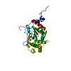
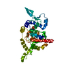
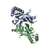
| Structure viewer | Molecule: MolmilJmol/JSmol |
|---|
- Downloads & links
-Download
| PDBx/mmCIF format | 4zx2.cif.gz | 92.2 KB | Display | PDBx/mmCIF format |
|---|---|---|---|---|
| PDB format | pdb4zx2.ent.gz | 66.2 KB | Display | PDB format |
| PDBx/mmJSON format | 4zx2.json.gz | Tree view | PDBx/mmJSON format | |
| Others |  Other downloads Other downloads |
-Validation report
| Arichive directory | https://data.pdbj.org/pub/pdb/validation_reports/zx/4zx2ftp://data.pdbj.org/pub/pdb/validation_reports/zx/4zx2 | HTTPS FTP |
|---|
-Related structure data
| Related structure data |  4zvzC 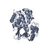 1s95S S: Starting model for refinement C: citing same article ( |
|---|---|
| Similar structure data |


-Links
 PDBj
PDBj


- Assembly
| Deposited unit | 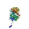
| ||||||||
|---|---|---|---|---|---|---|---|---|---|
| 1 |
| ||||||||
| Unit cell |
|
-Components
-Protein , 1 types, 1 molecules A
| #1: Protein | Mass: 37887.020 Da / Num. of mol.: 1 / Fragment: UNP residues 169-499 Source method: isolated from a genetically manipulated source Source: (gene. exp.) Homo sapiens (human) / Gene: PPP5C, PPP5 / Plasmid: pMal2cE / Production host:  References: UniProt: P53041, protein-serine/threonine phosphatase |
|---|
-Non-polymers , 5 types, 349 molecules 


| #2: Chemical |  Mass: 54.938 Da / Num. of mol.: 2 / Source method: obtained synthetically / Formula: Mn Mass: 54.938 Da / Num. of mol.: 2 / Source method: obtained synthetically / Formula: Mn#3: Chemical | ChemComp-4TE / ( |  Mass: 200.189 Da / Num. of mol.: 1 / Source method: obtained synthetically / Formula: C9H12O5 Mass: 200.189 Da / Num. of mol.: 1 / Source method: obtained synthetically / Formula: C9H12O5#4: Chemical | ChemComp-MPD / ( |  Mass: 118.174 Da / Num. of mol.: 1 / Source method: isolated from a natural source / Formula: C6H14O2 / Comment: precipitant*YM Mass: 118.174 Da / Num. of mol.: 1 / Source method: isolated from a natural source / Formula: C6H14O2 / Comment: precipitant*YM#5: Chemical | ChemComp-MRD / ( |  Mass: 118.174 Da / Num. of mol.: 1 / Source method: obtained synthetically / Formula: C6H14O2 / Comment: precipitant*YM Mass: 118.174 Da / Num. of mol.: 1 / Source method: obtained synthetically / Formula: C6H14O2 / Comment: precipitant*YM#6: Water | ChemComp-HOH / | Mass: 18.015 Da / Num. of mol.: 344 / Source method: isolated from a natural source / Formula: H2O |
|---|
-Experimental details
-Experiment
| Experiment | Method: X-RAY DIFFRACTION |
|---|
- Sample preparation
| Crystal | Density Matthews: 2.36 Å3/Da / Density % sol: 47.95 % |
|---|---|
| Crystal grow | Temperature: 289 K / Method: vapor diffusion, hanging drop / pH: 8 / Details: 25% MPD, 6% PEG, 10 mM Tris, pH 8.0 |
-Data collection
| Diffraction | Mean temperature: 200 K |
|---|---|
| Diffraction source | Source: SYNCHROTRON / Site: APS / Beamline: 24-ID-E / Wavelength: 0.97918 Å |
| Detector | Type: ADSC QUANTUM 315 / Detector: CCD / Date: Mar 5, 2014 |
| Radiation | Protocol: SINGLE WAVELENGTH / Monochromatic (M) / Laue (L): M / Scattering type: x-ray |
| Radiation wavelength | Wavelength: 0.97918 Å / Relative weight: 1 |
| Reflection | Resolution: 1.19→94.83 Å / Num. obs: 107757 / % possible obs: 94.8 % / Redundancy: 4.6 % / Rmerge(I) obs: 0.06 / Net I/σ(I): 18.3 |
| Reflection shell | Resolution: 1.19→1.25 Å / Redundancy: 3.3 % / Rmerge(I) obs: 0.594 / Mean I/σ(I) obs: 1.9 / % possible all: 72.9 |
- Processing
| Software |
| ||||||||||||||||||||||||||||||||||||||||||||||||||||||||||||||||||||||||||||||||||||||||||||||||||||||||||||||||||||||||||||||||||||||||||||||||||||||||||||||||||||||||||||||||||||||
|---|---|---|---|---|---|---|---|---|---|---|---|---|---|---|---|---|---|---|---|---|---|---|---|---|---|---|---|---|---|---|---|---|---|---|---|---|---|---|---|---|---|---|---|---|---|---|---|---|---|---|---|---|---|---|---|---|---|---|---|---|---|---|---|---|---|---|---|---|---|---|---|---|---|---|---|---|---|---|---|---|---|---|---|---|---|---|---|---|---|---|---|---|---|---|---|---|---|---|---|---|---|---|---|---|---|---|---|---|---|---|---|---|---|---|---|---|---|---|---|---|---|---|---|---|---|---|---|---|---|---|---|---|---|---|---|---|---|---|---|---|---|---|---|---|---|---|---|---|---|---|---|---|---|---|---|---|---|---|---|---|---|---|---|---|---|---|---|---|---|---|---|---|---|---|---|---|---|---|---|---|---|---|---|
| Refinement | Method to determine structure: MOLECULAR REPLACEMENT Starting model: PDB entry 1S95 Resolution: 1.23→40.96 Å / Cor.coef. Fo:Fc: 0.966 / Cor.coef. Fo:Fc free: 0.964 / SU B: 0.518 / SU ML: 0.023 / Cross valid method: THROUGHOUT / ESU R: 0.039 / ESU R Free: 0.04 / Stereochemistry target values: MAXIMUM LIKELIHOOD / Details: HYDROGENS HAVE BEEN USED IF PRESENT IN THE INPUT
| ||||||||||||||||||||||||||||||||||||||||||||||||||||||||||||||||||||||||||||||||||||||||||||||||||||||||||||||||||||||||||||||||||||||||||||||||||||||||||||||||||||||||||||||||||||||
| Solvent computation | Ion probe radii: 0.8 Å / Shrinkage radii: 0.8 Å / VDW probe radii: 1.2 Å / Solvent model: MASK | ||||||||||||||||||||||||||||||||||||||||||||||||||||||||||||||||||||||||||||||||||||||||||||||||||||||||||||||||||||||||||||||||||||||||||||||||||||||||||||||||||||||||||||||||||||||
| Displacement parameters | Biso mean: 10.248 Å2
| ||||||||||||||||||||||||||||||||||||||||||||||||||||||||||||||||||||||||||||||||||||||||||||||||||||||||||||||||||||||||||||||||||||||||||||||||||||||||||||||||||||||||||||||||||||||
| Refinement step | Cycle: LAST / Resolution: 1.23→40.96 Å
| ||||||||||||||||||||||||||||||||||||||||||||||||||||||||||||||||||||||||||||||||||||||||||||||||||||||||||||||||||||||||||||||||||||||||||||||||||||||||||||||||||||||||||||||||||||||
| Refine LS restraints |
|