 Movie
Movie Controller

[English] 日本語
 Yorodumi
- PDB-1xxo: X-ray crystal structure of mycobacterium tuberculosis pyridoxine ... -
+ Open data

- Basic information
| Entry | Database: PDB / ID: 1xxo | ||||||
|---|---|---|---|---|---|---|---|
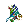
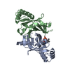
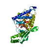
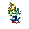
| Title | X-ray crystal structure of mycobacterium tuberculosis pyridoxine 5'-phosphate oxidase at 1.8 a resolution | ||||||
 Components Components | hypothetical protein Rv1155 | ||||||
 Keywords Keywords | OXIDOREDUCTASE / PYRIDOXINE 5'-PHOSPHATE / Structural Genomics / PSI / Protein Structure Initiative / TB Structural Genomics Consortium / TBSGC | ||||||
| Function / homology |  Function and homology information Function and homology informationvitamin B6 metabolic process / coenzyme F420 binding / oxidoreductase activity, acting on the CH-CH group of donors / Oxidoreductases / FMN binding / pyridoxal phosphate binding / protein homodimerization activity Similarity search - Function | ||||||
| Biological species |   Mycobacterium tuberculosis (bacteria) Mycobacterium tuberculosis (bacteria) | ||||||
| Method |  X-RAY DIFFRACTION / SYNCHROTRON / MAD / Resolution: 1.8 Å X-RAY DIFFRACTION / SYNCHROTRON / MAD / Resolution: 1.8 Å | ||||||
 Authors Authors | Biswal, B.K. / Cherney, M.M. / Wang, M. / Garen, C. / James, M.N.G. / TB Structural Genomics Consortium (TBSGC) | ||||||
 Citation Citation | Journal: To be Published Title: X-ray crystal structure of mycobacterium tuberculosis pyridoxine 5'-phosphate oxidase at 1.8 a resolution Authors: Biswal, B.K. / Cherney, M.M. / Wang, M. / Garen, C. / James, M.N.G. | ||||||
| History |
| ||||||
| Remark 400 | COMPOUND SUBUNIT A IN ENTRY 1XXO CORRESPOND TO SUBUNIT B IN 1Y30 AND 2AQ6 AND VICE VERSA |
- Structure visualization
| Structure viewer | Molecule: MolmilJmol/JSmol |
|---|
- Downloads & links
-Download
| PDBx/mmCIF format | 1xxo.cif.gz | 72.9 KB | Display | PDBx/mmCIF format |
|---|---|---|---|---|
| PDB format | pdb1xxo.ent.gz | 54.9 KB | Display | PDB format |
| PDBx/mmJSON format | 1xxo.json.gz | Tree view | PDBx/mmJSON format | |
| Others |  Other downloads Other downloads |
-Validation report
| Arichive directory | https://data.pdbj.org/pub/pdb/validation_reports/xx/1xxoftp://data.pdbj.org/pub/pdb/validation_reports/xx/1xxo | HTTPS FTP |
|---|
-Related structure data
| Related structure data | |
|---|---|
| Similar structure data | |
| Other databases |


-Links
 PDBj
PDBj- Assembly
| Deposited unit | 
| ||||||||
|---|---|---|---|---|---|---|---|---|---|
| 1 |
| ||||||||
| Unit cell |
|
-Components
| #1: Protein | Mass: 16321.480 Da / Num. of mol.: 2 Source method: isolated from a genetically manipulated source Source: (gene. exp.) Mycobacterium tuberculosis (bacteria) / Gene: RV1155 / Plasmid: PET / Species (production host): Escherichia coli / Production host: #2: Water | ChemComp-HOH / |  Mass: 18.015 Da / Num. of mol.: 323 / Source method: isolated from a natural source / Formula: H2O Mass: 18.015 Da / Num. of mol.: 323 / Source method: isolated from a natural source / Formula: H2O |
|---|
-Experimental details
-Experiment
| Experiment | Method: X-RAY DIFFRACTION |
|---|
- Sample preparation
| Crystal | Density Matthews: 2.1 Å3/Da / Density % sol: 41.4 % |
|---|---|
| Crystal grow | Temperature: 293 K / Method: vapor diffusion, hanging drop / pH: 8 Details: 12% PEG 8000, 8% ethylene glycerol, 25 mM potassium fluoride, pH 8.0, VAPOR DIFFUSION, HANGING DROP, temperature 293K |
-Data collection
| Diffraction | Mean temperature: 100 K | ||||||||||||
|---|---|---|---|---|---|---|---|---|---|---|---|---|---|
| Diffraction source | Source: SYNCHROTRON / Site: ALS  / Beamline: 8.3.1 / Wavelength: 1.339794, 1.105523, 1.033204 / Beamline: 8.3.1 / Wavelength: 1.339794, 1.105523, 1.033204 | ||||||||||||
| Detector | Type: ADSC QUANTUM 210 / Detector: CCD / Date: Feb 19, 2004 | ||||||||||||
| Radiation | Protocol: MAD / Monochromatic (M) / Laue (L): M / Scattering type: x-ray | ||||||||||||
| Radiation wavelength |
| ||||||||||||
| Reflection | Resolution: 1.8→40 Å / Num. obs: 22952 / % possible obs: 92.4 % / Observed criterion σ(F): 0 / Observed criterion σ(I): 0 / Redundancy: 2.1 % / Biso Wilson estimate: 11.4 Å2 / Rsym value: 0.063 / Net I/σ(I): 12.9 | ||||||||||||
| Reflection shell | Resolution: 1.8→1.9 Å / Redundancy: 1.4 % / Mean I/σ(I) obs: 2.2 / Num. unique all: 1699 / Rsym value: 0.271 |
- Processing
| Software |
| ||||||||||||||||||||||||||||||||||||||||||||||||||||||||||||
|---|---|---|---|---|---|---|---|---|---|---|---|---|---|---|---|---|---|---|---|---|---|---|---|---|---|---|---|---|---|---|---|---|---|---|---|---|---|---|---|---|---|---|---|---|---|---|---|---|---|---|---|---|---|---|---|---|---|---|---|---|---|
| Refinement | Method to determine structure: MAD / Resolution: 1.8→34.74 Å / Rfactor Rfree error: 0.005 / Data cutoff high absF: 1492756.07 / Data cutoff low absF: 0 / Isotropic thermal model: RESTRAINED / Cross valid method: THROUGHOUT / σ(F): 0 Details: INITIALLY THE STRUCTURE WAS SOLVED AT 2.2 A BY MAD METHOD WITH Ir AS THE ANOMALOUS SCATTERER. HIGH RESOLUTION DATA UP TO 1.8 A WAS COLLECTED LATER AND THE FINAL STRUCTURE WAS REFINED UP TO 1. ...Details: INITIALLY THE STRUCTURE WAS SOLVED AT 2.2 A BY MAD METHOD WITH Ir AS THE ANOMALOUS SCATTERER. HIGH RESOLUTION DATA UP TO 1.8 A WAS COLLECTED LATER AND THE FINAL STRUCTURE WAS REFINED UP TO 1.8 A. ELECTRON DENSITY WAS NOT OBSERVED FOR AMINO ACID RESIDUES 1-4 OF BOTH CHAINS.
| ||||||||||||||||||||||||||||||||||||||||||||||||||||||||||||
| Solvent computation | Solvent model: FLAT MODEL / Bsol: 59.1442 Å2 / ksol: 0.37374 e/Å3 | ||||||||||||||||||||||||||||||||||||||||||||||||||||||||||||
| Displacement parameters | Biso mean: 17.1 Å2
| ||||||||||||||||||||||||||||||||||||||||||||||||||||||||||||
| Refine analyze |
| ||||||||||||||||||||||||||||||||||||||||||||||||||||||||||||
| Refinement step | Cycle: LAST / Resolution: 1.8→34.74 Å
| ||||||||||||||||||||||||||||||||||||||||||||||||||||||||||||
| Refine LS restraints |
| ||||||||||||||||||||||||||||||||||||||||||||||||||||||||||||
| LS refinement shell | Resolution: 1.8→1.91 Å / Rfactor Rfree error: 0.017 / Total num. of bins used: 6
| ||||||||||||||||||||||||||||||||||||||||||||||||||||||||||||
| Xplor file |
|
