 Movie
Movie Controller

[English] 日本語
 Yorodumi
- PDB-1g76: X-RAY STRUCTURE OF ESCHERICHIA COLI PYRIDOXINE 5'-PHOSPHATE OXIDA... -
+ Open data

- Basic information
| Entry | Database: PDB / ID: 1g76 | ||||||
|---|---|---|---|---|---|---|---|
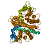
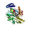
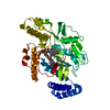
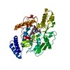
| Title | X-RAY STRUCTURE OF ESCHERICHIA COLI PYRIDOXINE 5'-PHOSPHATE OXIDASE COMPLEXED WITH PYRIDOXAL 5'-PHOSPHATE AT 2.0 A RESOLUTION | ||||||
 Components Components | PYRIDOXINE 5`-PHOSPHATE OXIDASE | ||||||
 Keywords Keywords | OXIDOREDUCTASE / PLP complex / FMN complex / pyridoxine 5'-phosphate | ||||||
| Function / homology |  Function and homology information Function and homology information'de novo' pyridoxal 5'-phosphate biosynthetic process / pyridoxal 5'-phosphate synthase / pyridoxamine phosphate oxidase activity / riboflavin binding / pyridoxal 5'-phosphate biosynthetic process / pyridoxine biosynthetic process / phosphate ion binding / FMN binding / pyridoxal phosphate binding / oxidoreductase activity ...'de novo' pyridoxal 5'-phosphate biosynthetic process / pyridoxal 5'-phosphate synthase / pyridoxamine phosphate oxidase activity / riboflavin binding / pyridoxal 5'-phosphate biosynthetic process / pyridoxine biosynthetic process / phosphate ion binding / FMN binding / pyridoxal phosphate binding / oxidoreductase activity / protein homodimerization activity / protein-containing complex / cytosol Similarity search - Function | ||||||
| Biological species |  | ||||||
| Method |  X-RAY DIFFRACTION / FOURIER SYNTHESIS / Resolution: 2.2 Å X-RAY DIFFRACTION / FOURIER SYNTHESIS / Resolution: 2.2 Å | ||||||
 Authors Authors | Safo, M.K. / Musayev, F.N. / di Salvo, M.L. / Schirch, V. | ||||||
 Citation Citation | Journal: J.Mol.Biol. / Year: 2001 Title: X-ray structure of Escherichia coli pyridoxine 5'-phosphate oxidase complexed with pyridoxal 5'-phosphate at 2.0 A resolution. Authors: Safo, M.K. / Musayev, F.N. / di Salvo, M.L. / Schirch, V. | ||||||
| History |
|
- Structure visualization
| Structure viewer | Molecule: MolmilJmol/JSmol |
|---|
- Downloads & links
-Download
| PDBx/mmCIF format | 1g76.cif.gz | 58.3 KB | Display | PDBx/mmCIF format |
|---|---|---|---|---|
| PDB format | pdb1g76.ent.gz | 41.3 KB | Display | PDB format |
| PDBx/mmJSON format | 1g76.json.gz | Tree view | PDBx/mmJSON format | |
| Others |  Other downloads Other downloads |
-Validation report
| Arichive directory | https://data.pdbj.org/pub/pdb/validation_reports/g7/1g76ftp://data.pdbj.org/pub/pdb/validation_reports/g7/1g76 | HTTPS FTP |
|---|
-Related structure data
| Related structure data |  1g77C  1g78C  1g79C 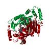 1dnlS S: Starting model for refinement C: citing same article ( |
|---|---|
| Similar structure data |


-Links
 PDBj
PDBj- Assembly
| Deposited unit | 
| ||||||||
|---|---|---|---|---|---|---|---|---|---|
| 1 | 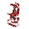
| ||||||||
| Unit cell |
| ||||||||
| Details | The biological assembly is a dimer, which was constructued from chain A and its symmetry partner generated by the two-fold axis |
-Components
| #1: Protein | Mass: 25585.188 Da / Num. of mol.: 1 Source method: isolated from a genetically manipulated source Source: (gene. exp.) References: UniProt: P28225, UniProt: P0AFI7*PLUS, pyridoxal 5'-phosphate synthase | ||||||
|---|---|---|---|---|---|---|---|
| #2: Chemical |   Mass: 94.971 Da / Num. of mol.: 2 / Source method: obtained synthetically / Formula: PO4 Mass: 94.971 Da / Num. of mol.: 2 / Source method: obtained synthetically / Formula: PO4#3: Chemical | ChemComp-FMN / |   Mass: 456.344 Da / Num. of mol.: 1 / Source method: obtained synthetically / Formula: C17H21N4O9P Mass: 456.344 Da / Num. of mol.: 1 / Source method: obtained synthetically / Formula: C17H21N4O9P#4: Chemical | ChemComp-PLP / |   Mass: 247.142 Da / Num. of mol.: 1 / Source method: obtained synthetically / Formula: C8H10NO6P Mass: 247.142 Da / Num. of mol.: 1 / Source method: obtained synthetically / Formula: C8H10NO6P#5: Water | ChemComp-HOH / |  Mass: 18.015 Da / Num. of mol.: 70 / Source method: isolated from a natural source / Formula: H2O Mass: 18.015 Da / Num. of mol.: 70 / Source method: isolated from a natural source / Formula: H2O |
-Experimental details
-Experiment
| Experiment | Method: X-RAY DIFFRACTION / Number of used crystals: 1 |
|---|
- Sample preparation
| Crystal | Density Matthews: 2.82 Å3/Da / Density % sol: 56.35 % | ||||||||||||||||||||
|---|---|---|---|---|---|---|---|---|---|---|---|---|---|---|---|---|---|---|---|---|---|
| Crystal grow | Temperature: 298 K / Method: vapor diffusion, hanging drop / pH: 7 Details: Ammonium sulfate and MES, pH 7.0, VAPOR DIFFUSION, HANGING DROP, temperature 298K | ||||||||||||||||||||
| Crystal grow | *PLUS pH: 6.2 / Details: macroseeding | ||||||||||||||||||||
| Components of the solutions | *PLUS
|
-Data collection
| Diffraction | Mean temperature: 98 K |
|---|---|
| Diffraction source | Source: ROTATING ANODE / Type: RIGAKU RU200 / Wavelength: 1.5418 Å |
| Detector | Type: RIGAKU RAXIS II / Detector: IMAGE PLATE / Date: Mar 29, 2000 / Details: mirrors |
| Radiation | Monochromator: MSC mirrors / Protocol: LAUE / Monochromatic (M) / Laue (L): L / Scattering type: neutron |
| Radiation wavelength | Wavelength: 1.5418 Å / Relative weight: 1 |
| Reflection | Resolution: 2.2→55 Å / Num. all: 14882 / Num. obs: 14882 / % possible obs: 95.9 % / Observed criterion σ(F): 0 / Observed criterion σ(I): 0 / Redundancy: 2.9 % / Biso Wilson estimate: 25.6 Å2 / Rmerge(I) obs: 0.055 / Rsym value: 5.8 / Net I/σ(I): 12.1 |
| Reflection shell | Resolution: 2.2→2.3 Å / Redundancy: 2.6 % / Rmerge(I) obs: 0.232 / Mean I/σ(I) obs: 2.2 / Num. unique all: 1772 / % possible all: 94.1 |
| Reflection | *PLUS Num. measured all: 43602 |
| Reflection shell | *PLUS % possible obs: 94.1 % / Num. possible: 4588 / Num. unique obs: 1772 |
- Processing
| Software |
| ||||||||||||||||||||||||||||||||||||
|---|---|---|---|---|---|---|---|---|---|---|---|---|---|---|---|---|---|---|---|---|---|---|---|---|---|---|---|---|---|---|---|---|---|---|---|---|---|
| Refinement | Method to determine structure: FOURIER SYNTHESIS Starting model: PDB entry 1DNL Resolution: 2.2→55 Å / Rfactor Rfree error: 0.01 / Data cutoff high absF: 87008.93 / Data cutoff low absF: 0 / Isotropic thermal model: RESTRAINED / Cross valid method: THROUGHOUT / σ(F): 1 / Stereochemistry target values: Engh and Huber / Details: bulk solvent correction
| ||||||||||||||||||||||||||||||||||||
| Solvent computation | Solvent model: FLAT MODEL / Bsol: 58.57 Å2 / ksol: 0.38 e/Å3 | ||||||||||||||||||||||||||||||||||||
| Displacement parameters | Biso mean: 45.4 Å2
| ||||||||||||||||||||||||||||||||||||
| Refine analyze |
| ||||||||||||||||||||||||||||||||||||
| Refinement step | Cycle: LAST / Resolution: 2.2→55 Å
| ||||||||||||||||||||||||||||||||||||
| Refine LS restraints |
| ||||||||||||||||||||||||||||||||||||
| LS refinement shell | Resolution: 2.2→2.34 Å / Rfactor Rfree error: 0.034 / Total num. of bins used: 6
| ||||||||||||||||||||||||||||||||||||
| Xplor file |
| ||||||||||||||||||||||||||||||||||||
| Software | *PLUS Name: CNS / Version: 0.5 / Classification: refinement | ||||||||||||||||||||||||||||||||||||
| Refine LS restraints | *PLUS
|
