 Movie
Movie Controller

[English] 日本語
 Yorodumi
- PDB-4zox: Crystal structure of the Saccharomyces cerevisiae Sqt1 bound to t... -
+ Open data

- Basic information
| Entry | Database: PDB / ID: 4zox | ||||||
|---|---|---|---|---|---|---|---|
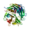
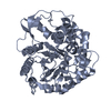
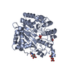
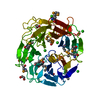
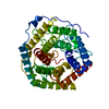
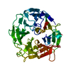
| Title | Crystal structure of the Saccharomyces cerevisiae Sqt1 bound to the N-terminus of the ribosomal protein L10 | ||||||
 Components Components |
| ||||||
 Keywords Keywords | CHAPERONE / ribosomal biogenesis / WD40 - repeat | ||||||
| Function / homology |  Function and homology information Function and homology informationSRP-dependent cotranslational protein targeting to membrane / GTP hydrolysis and joining of the 60S ribosomal subunit / Formation of a pool of free 40S subunits / Nonsense Mediated Decay (NMD) independent of the Exon Junction Complex (EJC) / Nonsense Mediated Decay (NMD) enhanced by the Exon Junction Complex (EJC) / L13a-mediated translational silencing of Ceruloplasmin expression / translational termination / ribosomal large subunit biogenesis / : / ribosomal large subunit assembly ...SRP-dependent cotranslational protein targeting to membrane / GTP hydrolysis and joining of the 60S ribosomal subunit / Formation of a pool of free 40S subunits / Nonsense Mediated Decay (NMD) independent of the Exon Junction Complex (EJC) / Nonsense Mediated Decay (NMD) enhanced by the Exon Junction Complex (EJC) / L13a-mediated translational silencing of Ceruloplasmin expression / translational termination / ribosomal large subunit biogenesis / : / ribosomal large subunit assembly / cytosolic large ribosomal subunit / cytoplasmic translation / structural constituent of ribosome / translation / cytosol / cytoplasm Similarity search - Function | ||||||
| Biological species |  | ||||||
| Method |  X-RAY DIFFRACTION / SYNCHROTRON / MOLECULAR REPLACEMENT / Resolution: 1.6 Å X-RAY DIFFRACTION / SYNCHROTRON / MOLECULAR REPLACEMENT / Resolution: 1.6 Å | ||||||
 Authors Authors | Pausch, P. / Altegoer, F. / Bange, G. | ||||||
 Citation Citation | Journal: Nat Commun / Year: 2015 Title: Co-translational capturing of nascent ribosomal proteins by their dedicated chaperones. Authors: Pausch, P. / Singh, U. / Ahmed, Y.L. / Pillet, B. / Murat, G. / Altegoer, F. / Stier, G. / Thoms, M. / Hurt, E. / Sinning, I. / Bange, G. / Kressler, D. | ||||||
| History |
|
- Structure visualization
| Structure viewer | Molecule: MolmilJmol/JSmol |
|---|
- Downloads & links
-Download
| PDBx/mmCIF format | 4zox.cif.gz | 103.2 KB | Display | PDBx/mmCIF format |
|---|---|---|---|---|
| PDB format | pdb4zox.ent.gz | 75.6 KB | Display | PDB format |
| PDBx/mmJSON format | 4zox.json.gz | Tree view | PDBx/mmJSON format | |
| Others |  Other downloads Other downloads |
-Validation report
| Arichive directory | https://data.pdbj.org/pub/pdb/validation_reports/zo/4zoxftp://data.pdbj.org/pub/pdb/validation_reports/zo/4zox | HTTPS FTP |
|---|
-Related structure data
| Related structure data |  4zn4C  4zovSC  4zoyC  4zozC S: Starting model for refinement C: citing same article ( |
|---|---|
| Similar structure data |


-Links
 PDBj
PDBj


- Assembly
| Deposited unit | 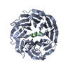
| |||||||||||||||
|---|---|---|---|---|---|---|---|---|---|---|---|---|---|---|---|---|
| 1 |
| |||||||||||||||
| Unit cell |
| |||||||||||||||
| Components on special symmetry positions |
|
-Components
| #1: Protein | Mass: 41408.727 Da / Num. of mol.: 1 / Fragment: UNP residues 53-431 Source method: isolated from a genetically manipulated source Source: (gene. exp.) Gene: SQT1, YIR012W, YIB12W / Plasmid: pETDuet-1 / Production host:  |
|---|---|
| #2: Protein/peptide | Mass: 3494.010 Da / Num. of mol.: 1 / Fragment: UNP residues 1-20 Source method: isolated from a genetically manipulated source Source: (gene. exp.) Gene: RPL10, GRC5, QSR1, YLR075W / Plasmid: pETDuet-1 / Production host: |
| #3: Water | ChemComp-HOH /  Mass: 18.015 Da / Num. of mol.: 559 / Source method: isolated from a natural source / Formula: H2O Mass: 18.015 Da / Num. of mol.: 559 / Source method: isolated from a natural source / Formula: H2O |
-Experimental details
-Experiment
| Experiment | Method: X-RAY DIFFRACTION |
|---|
- Sample preparation
| Crystal | Density Matthews: 1.95 Å3/Da / Density % sol: 36.76 % |
|---|---|
| Crystal grow | Temperature: 291 K / Method: vapor diffusion, sitting drop Details: 0.2 M Ca-acetate, 0.1 M Nacacodylate pH 6.5, 40% (v/v) PEG600 |
-Data collection
| Diffraction | Mean temperature: 100 K |
|---|---|
| Diffraction source | Source: SYNCHROTRON / Site: ESRF  / Beamline: ID29 / Wavelength: 0.97239 Å / Beamline: ID29 / Wavelength: 0.97239 Å |
| Detector | Type: PSI PILATUS 6M / Detector: PIXEL / Date: Apr 13, 2014 |
| Radiation | Protocol: SINGLE WAVELENGTH / Monochromatic (M) / Laue (L): M / Scattering type: x-ray |
| Radiation wavelength | Wavelength: 0.97239 Å / Relative weight: 1 |
| Reflection | Resolution: 1.6→44.63 Å / Num. obs: 44971 / % possible obs: 99.4 % / Redundancy: 4 % / Net I/σ(I): 18.5 |
- Processing
| Software |
| |||||||||||||||||||||||||||||||||||||||||||||||||||||||||||||||||||||||||||||||||||||||||||||||||||||||||||||||||||||||
|---|---|---|---|---|---|---|---|---|---|---|---|---|---|---|---|---|---|---|---|---|---|---|---|---|---|---|---|---|---|---|---|---|---|---|---|---|---|---|---|---|---|---|---|---|---|---|---|---|---|---|---|---|---|---|---|---|---|---|---|---|---|---|---|---|---|---|---|---|---|---|---|---|---|---|---|---|---|---|---|---|---|---|---|---|---|---|---|---|---|---|---|---|---|---|---|---|---|---|---|---|---|---|---|---|---|---|---|---|---|---|---|---|---|---|---|---|---|---|---|---|
| Refinement | Method to determine structure: MOLECULAR REPLACEMENT Starting model: 4ZOV Resolution: 1.6→44.626 Å / SU ML: 0.12 / Cross valid method: FREE R-VALUE / σ(F): 1.39 / Phase error: 16.62 / Stereochemistry target values: ML
| |||||||||||||||||||||||||||||||||||||||||||||||||||||||||||||||||||||||||||||||||||||||||||||||||||||||||||||||||||||||
| Solvent computation | Shrinkage radii: 0.9 Å / VDW probe radii: 1.11 Å / Solvent model: FLAT BULK SOLVENT MODEL | |||||||||||||||||||||||||||||||||||||||||||||||||||||||||||||||||||||||||||||||||||||||||||||||||||||||||||||||||||||||
| Refinement step | Cycle: LAST / Resolution: 1.6→44.626 Å
| |||||||||||||||||||||||||||||||||||||||||||||||||||||||||||||||||||||||||||||||||||||||||||||||||||||||||||||||||||||||
| Refine LS restraints |
| |||||||||||||||||||||||||||||||||||||||||||||||||||||||||||||||||||||||||||||||||||||||||||||||||||||||||||||||||||||||
| LS refinement shell |
|
