 Movie
Movie Controller

[English] 日本語
 Yorodumi
- PDB-4zow: Crystal structure of E. coli multidrug transporter MdfA in comple... -
+ Open data

- Basic information
| Entry | Database: PDB / ID: 4zow | ||||||
|---|---|---|---|---|---|---|---|
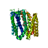
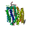
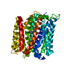
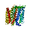
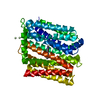
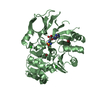
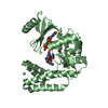
| Title | Crystal structure of E. coli multidrug transporter MdfA in complex with chloramphenicol | ||||||
 Components Components | Multidrug transporter MdfA | ||||||
 Keywords Keywords | TRANSPORT PROTEIN / MFS family / Multi-drug antiporter / MDFA / Cm | ||||||
| Function / homology |  Function and homology information Function and homology informationpotassium:proton antiporter activity / sodium:proton antiporter activity / xenobiotic detoxification by transmembrane export across the plasma membrane / : / Antimicrobial resistance / response to antibiotic / plasma membrane Similarity search - Function | ||||||
| Biological species |  | ||||||
| Method |  X-RAY DIFFRACTION / SYNCHROTRON / SAD / Resolution: 2.45 Å X-RAY DIFFRACTION / SYNCHROTRON / SAD / Resolution: 2.45 Å | ||||||
 Authors Authors | Zhang, X.C. / Heng, J. / Zhao, Y. / Wang, X. | ||||||
 Citation Citation | Journal: Cell Res. / Year: 2015 Title: Substrate-bound structure of the E. coli multidrug resistance transporter MdfA Authors: Heng, J. / Zhao, Y. / Liu, M. / Liu, Y. / Fan, J. / Wang, X. / Zhao, Y. / Zhang, X.C. | ||||||
| History |
|
- Structure visualization
| Structure viewer | Molecule: MolmilJmol/JSmol |
|---|
- Downloads & links
-Download
| PDBx/mmCIF format | 4zow.cif.gz | 88.3 KB | Display | PDBx/mmCIF format |
|---|---|---|---|---|
| PDB format | pdb4zow.ent.gz | 64.3 KB | Display | PDB format |
| PDBx/mmJSON format | 4zow.json.gz | Tree view | PDBx/mmJSON format | |
| Others |  Other downloads Other downloads |
-Validation report
| Arichive directory | https://data.pdbj.org/pub/pdb/validation_reports/zo/4zowftp://data.pdbj.org/pub/pdb/validation_reports/zo/4zow | HTTPS FTP |
|---|
-Related structure data


-Links
 PDBj
PDBj


- Assembly
| Deposited unit | 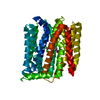
| ||||||||
|---|---|---|---|---|---|---|---|---|---|
| 1 |
| ||||||||
| Unit cell |
|
-Components
| #1: Protein | Mass: 42390.543 Da / Num. of mol.: 1 / Fragment: UNP residues 10-400 / Mutation: Q122R Source method: isolated from a genetically manipulated source Source: (gene. exp.) Strain: K12 / Gene: mdfA, cmlA, cmr, b0842, JW0826 Production host: References: UniProt: P0AEY8 |
|---|---|
| #2: Chemical | ChemComp-CLM /   Mass: 323.129 Da / Num. of mol.: 1 / Source method: obtained synthetically / Formula: C11H12Cl2N2O5 / Comment: antibiotic*YM Mass: 323.129 Da / Num. of mol.: 1 / Source method: obtained synthetically / Formula: C11H12Cl2N2O5 / Comment: antibiotic*YM |
| #3: Water | ChemComp-HOH /  Mass: 18.015 Da / Num. of mol.: 13 / Source method: isolated from a natural source / Formula: H2O Mass: 18.015 Da / Num. of mol.: 13 / Source method: isolated from a natural source / Formula: H2O |
-Experimental details
-Experiment
| Experiment | Method: X-RAY DIFFRACTION / Number of used crystals: 1 |
|---|
- Sample preparation
| Crystal | Density Matthews: 3.37 Å3/Da / Density % sol: 63.45 % |
|---|---|
| Crystal grow | Temperature: 295 K / Method: vapor diffusion, hanging drop / pH: 5.8 / Details: sodium acetate, PEG 400, magnesium acetate |
-Data collection
| Diffraction | Mean temperature: 100 K | ||||||||||||||||||||||||||||||||||||||||||||||||||||||||||||||||||
|---|---|---|---|---|---|---|---|---|---|---|---|---|---|---|---|---|---|---|---|---|---|---|---|---|---|---|---|---|---|---|---|---|---|---|---|---|---|---|---|---|---|---|---|---|---|---|---|---|---|---|---|---|---|---|---|---|---|---|---|---|---|---|---|---|---|---|---|
| Diffraction source | Source: SYNCHROTRON / Site: Photon Factory  / Beamline: BL-17A / Wavelength: 1 Å / Beamline: BL-17A / Wavelength: 1 Å | ||||||||||||||||||||||||||||||||||||||||||||||||||||||||||||||||||
| Detector | Type: DECTRIS PILATUS 6M / Detector: PIXEL / Date: May 7, 2014 | ||||||||||||||||||||||||||||||||||||||||||||||||||||||||||||||||||
| Radiation | Protocol: SINGLE WAVELENGTH / Monochromatic (M) / Laue (L): M / Scattering type: x-ray | ||||||||||||||||||||||||||||||||||||||||||||||||||||||||||||||||||
| Radiation wavelength | Wavelength: 1 Å / Relative weight: 1 | ||||||||||||||||||||||||||||||||||||||||||||||||||||||||||||||||||
| Reflection | Redundancy: 11.7 % / Number: 135599 / Rmerge(I) obs: 0.142 / Χ2: 1.03 / D res high: 3 Å / D res low: 50 Å / Num. obs: 11581 / % possible obs: 99.1 | ||||||||||||||||||||||||||||||||||||||||||||||||||||||||||||||||||
| Diffraction reflection shell |
| ||||||||||||||||||||||||||||||||||||||||||||||||||||||||||||||||||
| Reflection | Resolution: 2.45→50 Å / Num. obs: 21233 / % possible obs: 99.6 % / Redundancy: 6.4 % / Biso Wilson estimate: 49.78 Å2 / Rmerge(I) obs: 0.116 / Χ2: 1.023 / Net I/av σ(I): 16.882 / Net I/σ(I): 6.7 / Num. measured all: 136611 | ||||||||||||||||||||||||||||||||||||||||||||||||||||||||||||||||||
| Reflection shell | Diffraction-ID: 1 / Rejects: _
|
- Processing
| Software |
| |||||||||||||||||||||||||||||||||||||||||||||||||||||||||||||||
|---|---|---|---|---|---|---|---|---|---|---|---|---|---|---|---|---|---|---|---|---|---|---|---|---|---|---|---|---|---|---|---|---|---|---|---|---|---|---|---|---|---|---|---|---|---|---|---|---|---|---|---|---|---|---|---|---|---|---|---|---|---|---|---|---|
| Refinement | Method to determine structure: SAD / Resolution: 2.45→45.088 Å / FOM work R set: 0.8037 / SU ML: 0.3 / Cross valid method: FREE R-VALUE / σ(F): 1.34 / Phase error: 26.6 / Stereochemistry target values: ML
| |||||||||||||||||||||||||||||||||||||||||||||||||||||||||||||||
| Solvent computation | Shrinkage radii: 0.6 Å / VDW probe radii: 1.1 Å / Solvent model: FLAT BULK SOLVENT MODEL | |||||||||||||||||||||||||||||||||||||||||||||||||||||||||||||||
| Displacement parameters | Biso max: 125.94 Å2 / Biso mean: 53.39 Å2 / Biso min: 27.46 Å2 | |||||||||||||||||||||||||||||||||||||||||||||||||||||||||||||||
| Refinement step | Cycle: final / Resolution: 2.45→45.088 Å
| |||||||||||||||||||||||||||||||||||||||||||||||||||||||||||||||
| Refine LS restraints |
| |||||||||||||||||||||||||||||||||||||||||||||||||||||||||||||||
| LS refinement shell | Refine-ID: X-RAY DIFFRACTION / Total num. of bins used: 8
|
