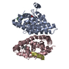
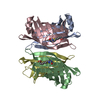
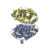
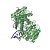
Thyroidhormonereceptorbeta / Nuclear receptor subfamily 1 group A member 2 / c-erbA-2 / c-erbA-beta
Mass: 28525.033 Da / Num. of mol.: 1 / Fragment: ligand binding domain unp residues 210-461 Source method: isolated from a genetically manipulated source Source: (gene. exp.) Homo sapiens (human) / Gene: THRB, ERBA2, NR1A2, THR1 / Production host: Escherichia coli (E. coli) / Strain (production host): BL21(DE3) / References: UniProt: P10828
#2: Protein/peptide
Nuclearreceptorcoactivator2 / NCoA-2 / Class E basic helix-loop-helix protein 75 / bHLHe75 / Transcriptional intermediary factor 2 / hTIF2
Mass: 1162.493 Da / Num. of mol.: 1 / Source method: obtained synthetically / Details: This sequence occurs naturally in humans / Source: (synth.) Homo sapiens (human) / References: UniProt: Q15596*PLUS
#3: Protein
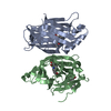
RetinoicacidreceptorRXR-alpha / Nuclear receptor subfamily 2 group B member 1 / Retinoid X receptor alpha
Mass: 25224.316 Da / Num. of mol.: 1 / Fragment: ligand binding domain, unp residues 231-455 / Source method: isolated from a natural source / Source: (natural) Homo sapiens (human) / References: UniProt: P19793
#4: Chemical
ChemComp-T3 / 3,5,3'TRIIODOTHYRONINE / T3 / THYROID HORMONE / LIOTHYRONINE
Mass: 650.973 Da / Num. of mol.: 1 / Source method: obtained synthetically / Formula: C15H12I3NO4
-
Experimental details
-
Experiment
Experiment
Method: X-RAY DIFFRACTION / Number of used crystals: 1
-
Sample preparation
Crystal
Density Matthews: 2.37 Å3/Da / Density % sol: 48.2 %
Crystal grow
Temperature: 294 K / Method: vapor diffusion, hanging drop / pH: 8 Details: 20% PEG 3350, 100 mM Tris pH 8.0, 0.1 M ammonium acetate, and 1 mM Methoprene acid
In the structure databanks used in Yorodumi, some data are registered as the other names, "COVID-19 virus" and "2019-nCoV". Here are the details of the virus and the list of structure data.
Jan 31, 2019. EMDB accession codes are about to change! (news from PDBe EMDB page)
EMDB accession codes are about to change! (news from PDBe EMDB page)
The allocation of 4 digits for EMDB accession codes will soon come to an end. Whilst these codes will remain in use, new EMDB accession codes will include an additional digit and will expand incrementally as the available range of codes is exhausted. The current 4-digit format prefixed with “EMD-” (i.e. EMD-XXXX) will advance to a 5-digit format (i.e. EMD-XXXXX), and so on. It is currently estimated that the 4-digit codes will be depleted around Spring 2019, at which point the 5-digit format will come into force.
The EM Navigator/Yorodumi systems omit the EMD- prefix.
Related info.:Q: What is EMD? / ID/Accession-code notation in Yorodumi/EM Navigator
Yorodumi is a browser for structure data from EMDB, PDB, SASBDB, etc.
This page is also the successor to EM Navigator detail page, and also detail information page/front-end page for Omokage search.
The word "yorodu" (or yorozu) is an old Japanese word meaning "ten thousand". "mi" (miru) is to see.
Related info.:EMDB / PDB / SASBDB / Comparison of 3 databanks / Yorodumi Search / Aug 31, 2016. New EM Navigator & Yorodumi / Yorodumi Papers / Jmol/JSmol / Function and homology information / Changes in new EM Navigator and Yorodumi
 Movie
Movie Controller

 Yorodumi
 Open data
 Basic information
 Components
 Keywords
 Function and homology information
 Homo sapiens (human)
 X-RAY DIFFRACTION /
  Authors
 Citation
 Structure visualization
 Downloads & links
 Other downloads
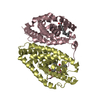


 PDBj
PDBj


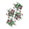
 Assembly

 Mass: 650.973 Da / Num. of mol.: 1 / Source method: obtained synthetically / Formula: C15H12I3NO4
 Sample preparation
 / Beamline: BL11-1 / Wavelength: 0.9 Å
 Processing