 Movie
Movie Controller


+ Open data

- Basic information
| Entry | Database: PDB / ID: 4zlx | ||||||
|---|---|---|---|---|---|---|---|
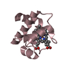
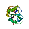
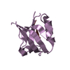
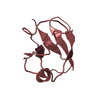
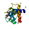
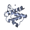
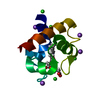
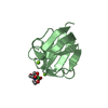
| Title | N-terminal DNA binding domain of the antitoxin Phd from phage P1 | ||||||
 Components Components | Antitoxin phd | ||||||
 Keywords Keywords | TRANSCRIPTION / transcription factor / toxin-antitoxin / DNA binding / intrinsic disorder / conditional cooperativity | ||||||
| Function / homology |  Function and homology information Function and homology informationtoxin sequestering activity / DNA-binding transcription repressor activity / protein-DNA complex / sequence-specific DNA binding / negative regulation of DNA-templated transcription / protein homodimerization activity / DNA binding Similarity search - Function | ||||||
| Biological species |  Enterobacteria phage P1 (virus) Enterobacteria phage P1 (virus) | ||||||
| Method |  X-RAY DIFFRACTION / MOLECULAR REPLACEMENT / Resolution: 2.31 Å X-RAY DIFFRACTION / MOLECULAR REPLACEMENT / Resolution: 2.31 Å | ||||||
 Authors Authors | Garcia-Pino, A. / Loris, R. | ||||||
 Citation Citation | Journal: Nat.Chem.Biol. / Year: 2016 Title: An intrinsically disordered entropic switch determines allostery in Phd-Doc regulation. Authors: Garcia-Pino, A. / De Gieter, S. / Talavera, A. / De Greve, H. / Efremov, R.G. / Loris, R. | ||||||
| History |
|
- Structure visualization
| Structure viewer | Molecule: MolmilJmol/JSmol |
|---|
- Downloads & links
-Download
| PDBx/mmCIF format | 4zlx.cif.gz | 52.1 KB | Display | PDBx/mmCIF format |
|---|---|---|---|---|
| PDB format | pdb4zlx.ent.gz | 36.8 KB | Display | PDB format |
| PDBx/mmJSON format | 4zlx.json.gz | Tree view | PDBx/mmJSON format | |
| Others |  Other downloads Other downloads |
-Validation report
| Arichive directory | https://data.pdbj.org/pub/pdb/validation_reports/zl/4zlxftp://data.pdbj.org/pub/pdb/validation_reports/zl/4zlx | HTTPS FTP |
|---|
-Related structure data
| Related structure data |  4zm0C  4zm2C  3hs2S S: Starting model for refinement C: citing same article ( |
|---|---|
| Similar structure data |
-Links
 PDBj
PDBj- Assembly
| Deposited unit | 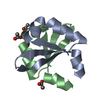
| ||||||||
|---|---|---|---|---|---|---|---|---|---|
| 1 |
| ||||||||
| Unit cell |
|
-Components
| #1: Protein | Mass: 6169.816 Da / Num. of mol.: 2 / Fragment: UNP residues 1-45 Source method: isolated from a genetically manipulated source Source: (gene. exp.) Enterobacteria phage P1 (virus) / Gene: phd / Production host:  #2: Chemical | ChemComp-ACT /   Mass: 59.044 Da / Num. of mol.: 5 / Source method: obtained synthetically / Formula: C2H3O2 Mass: 59.044 Da / Num. of mol.: 5 / Source method: obtained synthetically / Formula: C2H3O2#3: Water | ChemComp-HOH / |  Mass: 18.015 Da / Num. of mol.: 55 / Source method: isolated from a natural source / Formula: H2O Mass: 18.015 Da / Num. of mol.: 55 / Source method: isolated from a natural source / Formula: H2O |
|---|
-Experimental details
-Experiment
| Experiment | Method: X-RAY DIFFRACTION |
|---|
- Sample preparation
| Crystal | Density Matthews: 2.16 Å3/Da / Density % sol: 43.07 % |
|---|---|
| Crystal grow | Temperature: 293 K / Method: vapor diffusion, hanging drop Details: 200 mM Lithium Sulfate monohydrate, 20% w/v PEG3350 PH range: 6.0 - 7.0 |
-Data collection
| Diffraction | Mean temperature: 100 K |
|---|---|
| Diffraction source | Source: ROTATING ANODE / Type: RIGAKU MICROMAX-007 HF / Wavelength: 1.5418 Å |
| Detector | Type: RIGAKU SATURN 944+ / Detector: CCD / Date: Apr 19, 2012 |
| Radiation | Protocol: SINGLE WAVELENGTH / Monochromatic (M) / Laue (L): M / Scattering type: x-ray |
| Radiation wavelength | Wavelength: 1.5418 Å / Relative weight: 1 |
| Reflection | Resolution: 2.31→29.9 Å / Num. obs: 4518 / % possible obs: 97.3 % / Redundancy: 11.4 % / Biso Wilson estimate: 30.14 Å2 / Net I/σ(I): 57.8 |
| Reflection shell | Resolution: 2.31→2.39 Å / Redundancy: 8.8 % / Rmerge(I) obs: 0.13 / Mean I/σ(I) obs: 17.9 / % possible all: 86.7 |
- Processing
| Software |
| ||||||||||||||||||||||||||||||||||||||||||||||||||||||||||||||||||||||||||||||||||||||||||||||||||||||||||||||||||
|---|---|---|---|---|---|---|---|---|---|---|---|---|---|---|---|---|---|---|---|---|---|---|---|---|---|---|---|---|---|---|---|---|---|---|---|---|---|---|---|---|---|---|---|---|---|---|---|---|---|---|---|---|---|---|---|---|---|---|---|---|---|---|---|---|---|---|---|---|---|---|---|---|---|---|---|---|---|---|---|---|---|---|---|---|---|---|---|---|---|---|---|---|---|---|---|---|---|---|---|---|---|---|---|---|---|---|---|---|---|---|---|---|---|---|---|
| Refinement | Method to determine structure: MOLECULAR REPLACEMENT Starting model: 3HS2 Resolution: 2.31→29.86 Å / Cor.coef. Fo:Fc: 0.9335 / Cor.coef. Fo:Fc free: 0.8965 / SU R Cruickshank DPI: 0.329 / Cross valid method: THROUGHOUT / σ(F): 0 / SU R Blow DPI: 0.338 / SU Rfree Blow DPI: 0.24 / SU Rfree Cruickshank DPI: 0.241
| ||||||||||||||||||||||||||||||||||||||||||||||||||||||||||||||||||||||||||||||||||||||||||||||||||||||||||||||||||
| Displacement parameters | Biso mean: 32.74 Å2
| ||||||||||||||||||||||||||||||||||||||||||||||||||||||||||||||||||||||||||||||||||||||||||||||||||||||||||||||||||
| Refine analyze | Luzzati coordinate error obs: 0.302 Å | ||||||||||||||||||||||||||||||||||||||||||||||||||||||||||||||||||||||||||||||||||||||||||||||||||||||||||||||||||
| Refinement step | Cycle: LAST / Resolution: 2.31→29.86 Å
| ||||||||||||||||||||||||||||||||||||||||||||||||||||||||||||||||||||||||||||||||||||||||||||||||||||||||||||||||||
| Refine LS restraints |
| ||||||||||||||||||||||||||||||||||||||||||||||||||||||||||||||||||||||||||||||||||||||||||||||||||||||||||||||||||
| LS refinement shell | Resolution: 2.31→2.58 Å / Total num. of bins used: 5
| ||||||||||||||||||||||||||||||||||||||||||||||||||||||||||||||||||||||||||||||||||||||||||||||||||||||||||||||||||
| Refinement TLS params. | Method: refined / Refine-ID: X-RAY DIFFRACTION
| ||||||||||||||||||||||||||||||||||||||||||||||||||||||||||||||||||||||||||||||||||||||||||||||||||||||||||||||||||
| Refinement TLS group |
|
