 Movie
Movie Controller

[English] 日本語
 Yorodumi
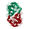
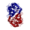
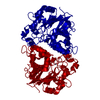
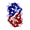
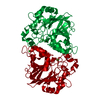
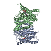
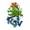
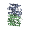
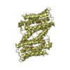
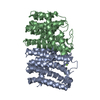
- PDB-4z67: The 1.5-angstrom crystal structure of Mn(2+)-bound PqqB from Pseu... -
+ Open data

- Basic information
| Entry | Database: PDB / ID: 4z67 | ||||||
|---|---|---|---|---|---|---|---|
| Title | The 1.5-angstrom crystal structure of Mn(2+)-bound PqqB from Pseudomonas Putida | ||||||
 Components Components | Coenzyme PQQ synthesis protein B | ||||||
 Keywords Keywords | HORMONE / PqqB / PQQ / metallo-beta-lactamase / beta-lactamase / Pyrroloquinoline quinone | ||||||
| Function / homology |  Function and homology information Function and homology information | ||||||
| Biological species |  Pseudomonas putida (bacteria) Pseudomonas putida (bacteria) | ||||||
| Method |  X-RAY DIFFRACTION / SYNCHROTRON / MOLECULAR REPLACEMENT / molecular replacement / Resolution: 1.5 Å X-RAY DIFFRACTION / SYNCHROTRON / MOLECULAR REPLACEMENT / molecular replacement / Resolution: 1.5 Å | ||||||
 Authors Authors | Tu, X. / Wilmot, C.M. | ||||||
 Citation Citation | Journal: To Be Published Title: Crystal structures reveal metal-binding plasticity at the active site of PqqB Authors: Tu, X. / Wilmot, C.M. | ||||||
| History |
|
- Structure visualization
| Structure viewer | Molecule: MolmilJmol/JSmol |
|---|
- Downloads & links
-Download
| PDBx/mmCIF format | 4z67.cif.gz | 153.4 KB | Display | PDBx/mmCIF format |
|---|---|---|---|---|
| PDB format | pdb4z67.ent.gz | 120 KB | Display | PDB format |
| PDBx/mmJSON format | 4z67.json.gz | Tree view | PDBx/mmJSON format | |
| Others |  Other downloads Other downloads |
-Validation report
| Arichive directory | https://data.pdbj.org/pub/pdb/validation_reports/z6/4z67ftp://data.pdbj.org/pub/pdb/validation_reports/z6/4z67 | HTTPS FTP |
|---|
-Related structure data


-Links
 PDBj
PDBj


- Assembly
| Deposited unit | 
| |||||||||
|---|---|---|---|---|---|---|---|---|---|---|
| 1 | 
| |||||||||
| Unit cell |
| |||||||||
| Components on special symmetry positions |
|
-Components
-Protein , 1 types, 1 molecules A
| #1: Protein | Mass: 34123.656 Da / Num. of mol.: 1 Source method: isolated from a genetically manipulated source Source: (gene. exp.) Pseudomonas putida (strain KT2440) (bacteria)Strain: KT2440 / Gene: pqqB, PP_0379 / Production host: |
|---|
-Non-polymers , 5 types, 322 molecules 


| #2: Chemical | ChemComp-ZN /  Mass: 65.409 Da / Num. of mol.: 1 / Source method: obtained synthetically / Formula: Zn Mass: 65.409 Da / Num. of mol.: 1 / Source method: obtained synthetically / Formula: Zn |
|---|---|
| #3: Chemical | ChemComp-MLT /  Mass: 134.087 Da / Num. of mol.: 1 / Source method: obtained synthetically / Formula: C4H6O5 Mass: 134.087 Da / Num. of mol.: 1 / Source method: obtained synthetically / Formula: C4H6O5 |
| #4: Chemical | ChemComp-MN /  Mass: 54.938 Da / Num. of mol.: 1 / Source method: obtained synthetically / Formula: Mn Mass: 54.938 Da / Num. of mol.: 1 / Source method: obtained synthetically / Formula: Mn |
| #5: Chemical | ChemComp-NA /  Mass: 22.990 Da / Num. of mol.: 1 / Source method: obtained synthetically / Formula: Na Mass: 22.990 Da / Num. of mol.: 1 / Source method: obtained synthetically / Formula: Na |
| #6: Water | ChemComp-HOH / Mass: 18.015 Da / Num. of mol.: 318 / Source method: isolated from a natural source / Formula: H2O |
-Experimental details
-Experiment
| Experiment | Method: X-RAY DIFFRACTION / Number of used crystals: 1 |
|---|
- Sample preparation
| Crystal | Density Matthews: 2.92 Å3/Da / Density % sol: 57.92 % |
|---|---|
| Crystal grow | Temperature: 298 K / Method: vapor diffusion, hanging drop / pH: 6.7 Details: 11% PEG4000, 0.2M NaCl, 1 mM MnCl2, 1 mM Malic acid, 0.1M Bis-Tris Propane pH6.7 |
-Data collection
| Diffraction | Mean temperature: 108 K | |||||||||||||||||||||||||||
|---|---|---|---|---|---|---|---|---|---|---|---|---|---|---|---|---|---|---|---|---|---|---|---|---|---|---|---|---|
| Diffraction source | Source: SYNCHROTRON / Site: APS  / Beamline: 23-ID-D / Wavelength: 0.987 Å / Beamline: 23-ID-D / Wavelength: 0.987 Å | |||||||||||||||||||||||||||
| Detector | Type: DECTRIS PILATUS 6M / Detector: PIXEL / Date: Feb 22, 2014 | |||||||||||||||||||||||||||
| Radiation | Protocol: SINGLE WAVELENGTH / Monochromatic (M) / Laue (L): M / Scattering type: x-ray | |||||||||||||||||||||||||||
| Radiation wavelength | Wavelength: 0.987 Å / Relative weight: 1 | |||||||||||||||||||||||||||
| Reflection | Resolution: 1.5→29.35 Å / Num. obs: 65627 / % possible obs: 99.7 % / Redundancy: 8.6 % / Biso Wilson estimate: 19.86 Å2 / CC1/2: 0.999 / Rmerge(I) obs: 0.065 / Rpim(I) all: 0.023 / Net I/σ(I): 19 / Num. measured all: 567404 | |||||||||||||||||||||||||||
| Reflection shell | Diffraction-ID: 1 / Rejects: _
|
-Phasing
| Phasing | Method: molecular replacement |
|---|
- Processing
| Software |
| |||||||||||||||||||||||||||||||||||||||||||||||||||||||||||||||||||||||||||||||||||||||||||||||||||||||||||||||||||||||||||||||||||||||||||||||||||||||||||||||||||||||||||||||
|---|---|---|---|---|---|---|---|---|---|---|---|---|---|---|---|---|---|---|---|---|---|---|---|---|---|---|---|---|---|---|---|---|---|---|---|---|---|---|---|---|---|---|---|---|---|---|---|---|---|---|---|---|---|---|---|---|---|---|---|---|---|---|---|---|---|---|---|---|---|---|---|---|---|---|---|---|---|---|---|---|---|---|---|---|---|---|---|---|---|---|---|---|---|---|---|---|---|---|---|---|---|---|---|---|---|---|---|---|---|---|---|---|---|---|---|---|---|---|---|---|---|---|---|---|---|---|---|---|---|---|---|---|---|---|---|---|---|---|---|---|---|---|---|---|---|---|---|---|---|---|---|---|---|---|---|---|---|---|---|---|---|---|---|---|---|---|---|---|---|---|---|---|---|---|---|---|
| Refinement | Method to determine structure: MOLECULAR REPLACEMENT / Resolution: 1.5→29.35 Å / FOM work R set: 0.9072 / SU ML: 0.2 / Cross valid method: FREE R-VALUE / σ(F): 1.36 / Phase error: 15.5 / Stereochemistry target values: ML
| |||||||||||||||||||||||||||||||||||||||||||||||||||||||||||||||||||||||||||||||||||||||||||||||||||||||||||||||||||||||||||||||||||||||||||||||||||||||||||||||||||||||||||||||
| Solvent computation | Shrinkage radii: 0.9 Å / VDW probe radii: 1.11 Å / Solvent model: FLAT BULK SOLVENT MODEL | |||||||||||||||||||||||||||||||||||||||||||||||||||||||||||||||||||||||||||||||||||||||||||||||||||||||||||||||||||||||||||||||||||||||||||||||||||||||||||||||||||||||||||||||
| Displacement parameters | Biso max: 66.42 Å2 / Biso mean: 26.45 Å2 / Biso min: 11.73 Å2 | |||||||||||||||||||||||||||||||||||||||||||||||||||||||||||||||||||||||||||||||||||||||||||||||||||||||||||||||||||||||||||||||||||||||||||||||||||||||||||||||||||||||||||||||
| Refinement step | Cycle: final / Resolution: 1.5→29.35 Å
| |||||||||||||||||||||||||||||||||||||||||||||||||||||||||||||||||||||||||||||||||||||||||||||||||||||||||||||||||||||||||||||||||||||||||||||||||||||||||||||||||||||||||||||||
| Refine LS restraints |
| |||||||||||||||||||||||||||||||||||||||||||||||||||||||||||||||||||||||||||||||||||||||||||||||||||||||||||||||||||||||||||||||||||||||||||||||||||||||||||||||||||||||||||||||
| LS refinement shell | Refine-ID: X-RAY DIFFRACTION / Total num. of bins used: 24
|
