| Entry | Database: PDB / ID: 4z39
|
|---|
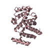
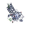
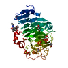
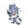
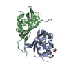
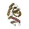
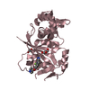
| Title | Structure of OBP3 from the vetch aphid Megoura viciae |
|---|
 Components Components | Odorant-binding protein |
|---|
 Keywords Keywords | odorant binding protein |
|---|
| Function / homology | Pheromone/general odorant binding protein / PBP/GOBP family / Pheromone/general odorant binding protein superfamily / odorant binding / sensory perception of smell / : / membrane / Odorant binding protein 3 / Odorant-binding protein Function and homology information Function and homology information |
|---|
| Biological species |  Megoura viciae (vetch aphid) Megoura viciae (vetch aphid) |
|---|
| Method |  X-RAY DIFFRACTION / SYNCHROTRON / MOLECULAR REPLACEMENT / Resolution: 1.3 Å X-RAY DIFFRACTION / SYNCHROTRON / MOLECULAR REPLACEMENT / Resolution: 1.3 Å |
|---|
 Authors Authors | Northey, T. / Venthur, H. / De Biasio, F. / Chauviac, F.-X. / Cole, A.R. / Field, L.M. / Zhou, J.-J. / Keep, N.H. |
|---|
| Funding support |  United Kingdom, 2items United Kingdom, 2items | Organization | Grant number | Country |
|---|
| Biotechnology and Biological Sciences Research Council | BB/L001683/1 | United Kingdom | | Biotechnology and Biological Sciences Research Council | BB/I024941/1 | United Kingdom |
|
|---|
 Citation Citation | Journal: Sci Rep / Year: 2016
Title: Crystal Structures and Binding Dynamics of Odorant-Binding Protein 3 from two aphid species Megoura viciae and Nasonovia ribisnigri.
Authors: Northey, T. / Venthur, H. / De Biasio, F. / Chauviac, F.X. / Cole, A. / Ribeiro, K.A. / Grossi, G. / Falabella, P. / Field, L.M. / Keep, N.H. / Zhou, J.J. |
|---|
| History | | Deposition | Mar 31, 2015 | Deposition site: RCSB / Processing site: PDBE |
|---|
| Revision 1.0 | Apr 13, 2016 | Provider: repository / Type: Initial release |
|---|
| Revision 1.1 | Apr 20, 2016 | Group: Database references |
|---|
| Revision 1.2 | Aug 30, 2017 | Group: Author supporting evidence / Category: pdbx_audit_support / Item: _pdbx_audit_support.funding_organization |
|---|
| Revision 1.3 | May 1, 2024 | Group: Data collection / Database references / Refinement description
Category: chem_comp_atom / chem_comp_bond ...chem_comp_atom / chem_comp_bond / database_2 / pdbx_initial_refinement_model / struct_ncs_dom_lim
Item: _database_2.pdbx_DOI / _database_2.pdbx_database_accession ..._database_2.pdbx_DOI / _database_2.pdbx_database_accession / _struct_ncs_dom_lim.beg_auth_comp_id / _struct_ncs_dom_lim.beg_label_asym_id / _struct_ncs_dom_lim.beg_label_comp_id / _struct_ncs_dom_lim.beg_label_seq_id / _struct_ncs_dom_lim.end_auth_comp_id / _struct_ncs_dom_lim.end_label_asym_id / _struct_ncs_dom_lim.end_label_comp_id / _struct_ncs_dom_lim.end_label_seq_id |
|---|
| Revision 1.4 | Oct 23, 2024 | Group: Structure summary / Category: pdbx_entry_details / pdbx_modification_feature |
|---|
|
|---|
 Movie
Movie Controller


 Open data
 Basic information
 Structure visualization
 Downloads & links
 Other downloads
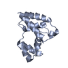


 PDBj
PDBj Assembly
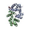


 Mass: 92.094 Da / Num. of mol.: 2 / Source method: obtained synthetically / Formula: C3H8O3
 Mass: 96.063 Da / Num. of mol.: 2 / Source method: obtained synthetically / Formula: SO4
 Mass: 18.015 Da / Num. of mol.: 199 / Source method: isolated from a natural source / Formula: H2O
 Sample preparation
 Processing