 Movie
Movie Controller


+ Open data

- Basic information
| Entry | Database: PDB / ID: 4yxk | ||||||
|---|---|---|---|---|---|---|---|
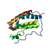
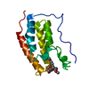
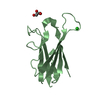
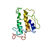
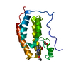
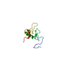
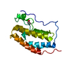
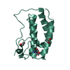
| Title | Crystal structure of Elk prion protein complexed with POM1 FAB | ||||||
 Components Components |
| ||||||
 Keywords Keywords | IMMUNE SYSTEM / PRION / ANTIBODY / Immune system complex | ||||||
| Function / homology |  Function and homology information Function and homology informationside of membrane / protein homooligomerization / copper ion binding / Golgi apparatus / plasma membrane Similarity search - Function | ||||||
| Biological species |  | ||||||
| Method |  X-RAY DIFFRACTION / SYNCHROTRON / MOLECULAR REPLACEMENT / Resolution: 2.805 Å X-RAY DIFFRACTION / SYNCHROTRON / MOLECULAR REPLACEMENT / Resolution: 2.805 Å | ||||||
 Authors Authors | Baral, P.K. / Swayampakula, M. / James, M.N.G. | ||||||
| Funding support |  Canada, 1items Canada, 1items
| ||||||
 Citation Citation | Journal: J.Struct.Biol. / Year: 2015 Title: X-ray structural and molecular dynamical studies of the globular domains of cow, deer, elk and Syrian hamster prion proteins. Authors: Baral, P.K. / Swayampakula, M. / Aguzzi, A. / James, M.N. | ||||||
| History |
|
- Structure visualization
| Structure viewer | Molecule: MolmilJmol/JSmol |
|---|
- Downloads & links
-Download
| PDBx/mmCIF format | 4yxk.cif.gz | 120.4 KB | Display | PDBx/mmCIF format |
|---|---|---|---|---|
| PDB format | pdb4yxk.ent.gz | 91.5 KB | Display | PDB format |
| PDBx/mmJSON format | 4yxk.json.gz | Tree view | PDBx/mmJSON format | |
| Others |  Other downloads Other downloads |
-Validation report
| Summary document | 4yxk_validation.pdf.gz | 451.1 KB | Display | wwPDB validaton report |
|---|---|---|---|---|
| Full document | 4yxk_full_validation.pdf.gz | 490.9 KB | Display | |
| Data in XML | 4yxk_validation.xml.gz | 25.8 KB | Display | |
| Data in CIF | 4yxk_validation.cif.gz | 35.1 KB | Display | |
| Arichive directory | https://data.pdbj.org/pub/pdb/validation_reports/yx/4yxkftp://data.pdbj.org/pub/pdb/validation_reports/yx/4yxk | HTTPS FTP |
-Related structure data
| Related structure data |  4yx2C  4yxhC 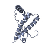 4yxlC  4dgiS  4h88S C: citing same article ( S: Starting model for refinement |
|---|---|
| Similar structure data |


-Links
 PDBj
PDBj


- Assembly
| Deposited unit | 
| ||||||||
|---|---|---|---|---|---|---|---|---|---|
| 1 | 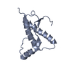
| ||||||||
| 2 | 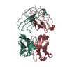
| ||||||||
| Unit cell |
|
-Components
| #1: Protein | Mass: 15931.748 Da / Num. of mol.: 1 Source method: isolated from a genetically manipulated source Source: (gene. exp.)  |
|---|---|
| #2: Antibody | Mass: 23397.078 Da / Num. of mol.: 1 Source method: isolated from a genetically manipulated source Source: (gene. exp.) |
| #3: Antibody | Mass: 23509.701 Da / Num. of mol.: 1 Source method: isolated from a genetically manipulated source Source: (gene. exp.) |
| #4: Chemical | ChemComp-NA /   Mass: 22.990 Da / Num. of mol.: 1 / Source method: obtained synthetically / Formula: Na Mass: 22.990 Da / Num. of mol.: 1 / Source method: obtained synthetically / Formula: Na |
| #5: Water | ChemComp-HOH /  Mass: 18.015 Da / Num. of mol.: 73 / Source method: isolated from a natural source / Formula: H2O Mass: 18.015 Da / Num. of mol.: 73 / Source method: isolated from a natural source / Formula: H2O |
| Has protein modification | Y |
-Experimental details
-Experiment
| Experiment | Method: X-RAY DIFFRACTION |
|---|
- Sample preparation
| Crystal | Density Matthews: 2.8 Å3/Da / Density % sol: 56.08 % |
|---|---|
| Crystal grow | Temperature: 295 K / Method: vapor diffusion, sitting drop Details: 25% PEG3350, 0.1 M BIS-TRIS, 0.2 M lithium sulphate |
-Data collection
| Diffraction | Mean temperature: 100 K |
|---|---|
| Diffraction source | Source: SYNCHROTRON / Site: SSRL  / Beamline: BL9-1 / Wavelength: 0.97945 Å / Beamline: BL9-1 / Wavelength: 0.97945 Å |
| Detector | Type: PSI PILATUS 6M / Detector: PIXEL / Date: Mar 31, 2013 |
| Radiation | Protocol: SINGLE WAVELENGTH / Monochromatic (M) / Laue (L): M / Scattering type: x-ray |
| Radiation wavelength | Wavelength: 0.97945 Å / Relative weight: 1 |
| Reflection | Resolution: 2.8→50 Å / Num. obs: 15989 / % possible obs: 98.4 % / Redundancy: 3.9 % / Rsym value: 0.11 / Net I/σ(I): 10.2 |
| Reflection shell | Resolution: 2.8→2.9 Å / Redundancy: 3.8 % / Mean I/σ(I) obs: 1.4 / % possible all: 99.3 |
- Processing
| Software |
| |||||||||||||||||||||||||||||||||||||||||||||||||
|---|---|---|---|---|---|---|---|---|---|---|---|---|---|---|---|---|---|---|---|---|---|---|---|---|---|---|---|---|---|---|---|---|---|---|---|---|---|---|---|---|---|---|---|---|---|---|---|---|---|---|
| Refinement | Method to determine structure: MOLECULAR REPLACEMENT Starting model: 4H88, 4DGI Resolution: 2.805→42.161 Å / SU ML: 0.44 / Cross valid method: FREE R-VALUE / σ(F): 1.35 / Phase error: 37.72 / Stereochemistry target values: ML
| |||||||||||||||||||||||||||||||||||||||||||||||||
| Solvent computation | Shrinkage radii: 0.9 Å / VDW probe radii: 1.11 Å / Solvent model: FLAT BULK SOLVENT MODEL | |||||||||||||||||||||||||||||||||||||||||||||||||
| Refinement step | Cycle: LAST / Resolution: 2.805→42.161 Å
| |||||||||||||||||||||||||||||||||||||||||||||||||
| Refine LS restraints |
| |||||||||||||||||||||||||||||||||||||||||||||||||
| LS refinement shell |
|
