 Movie
Movie Controller

[English] 日本語
 Yorodumi
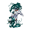
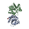
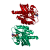
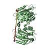
- PDB-4ygi: Crystal Structure of SUVH5 SRA bound to fully hydroxymethylated CG DNA -
+ Open data

- Basic information
| Entry | Database: PDB / ID: 4ygi | ||||||
|---|---|---|---|---|---|---|---|
| Title | Crystal Structure of SUVH5 SRA bound to fully hydroxymethylated CG DNA | ||||||
 Components Components |
| ||||||
 Keywords Keywords | TRANSFERASE/DNA / SUVH5 SRA / Fully hydroxymethylated CG / 5-hydroxymethylcytosine / 5hmC binding protein. / TRANSFERASE-DNA complex | ||||||
| Function / homology |  Function and homology information Function and homology information[histone H3]-lysine9 N-methyltransferase / histone H3K9 monomethyltransferase activity / double-stranded methylated DNA binding / regulatory ncRNA-mediated heterochromatin formation / histone methyltransferase activity / histone acetyltransferase complex / chromosome, centromeric region / epigenetic regulation of gene expression / Transferases; Transferring one-carbon groups; Methyltransferases / double-stranded DNA binding ...[histone H3]-lysine9 N-methyltransferase / histone H3K9 monomethyltransferase activity / double-stranded methylated DNA binding / regulatory ncRNA-mediated heterochromatin formation / histone methyltransferase activity / histone acetyltransferase complex / chromosome, centromeric region / epigenetic regulation of gene expression / Transferases; Transferring one-carbon groups; Methyltransferases / double-stranded DNA binding / methylation / zinc ion binding / nucleus Similarity search - Function | ||||||
| Biological species |  synthetic construct (others) | ||||||
| Method |  X-RAY DIFFRACTION / SYNCHROTRON / MOLECULAR REPLACEMENT / molecular replacement / Resolution: 2.6 Å X-RAY DIFFRACTION / SYNCHROTRON / MOLECULAR REPLACEMENT / molecular replacement / Resolution: 2.6 Å | ||||||
 Authors Authors | Rajakumara, E. | ||||||
 Citation Citation | Journal: Sci Rep / Year: 2016 Title: Mechanistic insights into the recognition of 5-methylcytosine oxidation derivatives by the SUVH5 SRA domain Authors: Rajakumara, E. / Nakarakanti, N.K. / Nivya, M.A. / Satish, M. | ||||||
| History |
|
- Structure visualization
| Structure viewer | Molecule: MolmilJmol/JSmol |
|---|
- Downloads & links
-Download
| PDBx/mmCIF format | 4ygi.cif.gz | 52.8 KB | Display | PDBx/mmCIF format |
|---|---|---|---|---|
| PDB format | pdb4ygi.ent.gz | 33.3 KB | Display | PDB format |
| PDBx/mmJSON format | 4ygi.json.gz | Tree view | PDBx/mmJSON format | |
| Others |  Other downloads Other downloads |
-Validation report
| Arichive directory | https://data.pdbj.org/pub/pdb/validation_reports/yg/4ygiftp://data.pdbj.org/pub/pdb/validation_reports/yg/4ygi | HTTPS FTP |
|---|
-Related structure data
| Related structure data |  3q0bS S: Starting model for refinement |
|---|---|
| Similar structure data |


-Links
 PDBj
PDBj


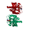
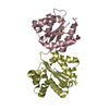
- Assembly
| Deposited unit | 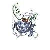
| ||||||||
|---|---|---|---|---|---|---|---|---|---|
| 1 | 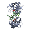
| ||||||||
| Unit cell |
|
-Components
| #1: Protein | Mass: 18515.920 Da / Num. of mol.: 1 / Fragment: SUVH5 SRA DOMAIN, UNP residues 362-528 Source method: isolated from a genetically manipulated source Source: (gene. exp.)  References: UniProt: O82175, histone-lysine N-methyltransferase | ||
|---|---|---|---|
| #2: DNA chain | Mass: 3378.235 Da / Num. of mol.: 1 / Source method: obtained synthetically / Details: CHEMICALLY SYNTHESIZED / Source: (synth.) synthetic construct (others) | ||
| #3: Chemical |   Mass: 24.305 Da / Num. of mol.: 3 / Source method: obtained synthetically / Formula: Mg Mass: 24.305 Da / Num. of mol.: 3 / Source method: obtained synthetically / Formula: Mg#4: Water | ChemComp-HOH / |  Mass: 18.015 Da / Num. of mol.: 16 / Source method: isolated from a natural source / Formula: H2O Mass: 18.015 Da / Num. of mol.: 16 / Source method: isolated from a natural source / Formula: H2O |
-Experimental details
-Experiment
| Experiment | Method: X-RAY DIFFRACTION / Number of used crystals: 1 |
|---|
- Sample preparation
| Crystal | Density Matthews: 2.46 Å3/Da / Density % sol: 49.96 % |
|---|---|
| Crystal grow | Temperature: 291 K / Method: vapor diffusion, sitting drop / pH: 6.5 Details: 0.2M Sodium chloride, 0.1M BIS-TRIS pH 6.5, 25%(w/v) Polyethylene glycol 3,350 |
-Data collection
| Diffraction | Mean temperature: 100 K | |||||||||||||||||||||||||||||||||||||||||||||||||||||||||||||||||||||||||||||
|---|---|---|---|---|---|---|---|---|---|---|---|---|---|---|---|---|---|---|---|---|---|---|---|---|---|---|---|---|---|---|---|---|---|---|---|---|---|---|---|---|---|---|---|---|---|---|---|---|---|---|---|---|---|---|---|---|---|---|---|---|---|---|---|---|---|---|---|---|---|---|---|---|---|---|---|---|---|---|
| Diffraction source | Source: SYNCHROTRON / Site: NSLS  / Beamline: X29A / Wavelength: 1.0718 Å / Beamline: X29A / Wavelength: 1.0718 Å | |||||||||||||||||||||||||||||||||||||||||||||||||||||||||||||||||||||||||||||
| Detector | Type: ADSC QUANTUM 315r / Detector: CCD / Date: Nov 8, 2011 | |||||||||||||||||||||||||||||||||||||||||||||||||||||||||||||||||||||||||||||
| Radiation | Monochromator: Si(111) / Protocol: SINGLE WAVELENGTH / Monochromatic (M) / Laue (L): M / Scattering type: x-ray | |||||||||||||||||||||||||||||||||||||||||||||||||||||||||||||||||||||||||||||
| Radiation wavelength | Wavelength: 1.0718 Å / Relative weight: 1 | |||||||||||||||||||||||||||||||||||||||||||||||||||||||||||||||||||||||||||||
| Reflection | Resolution: 2.6→50 Å / Num. obs: 7060 / % possible obs: 98.8 % / Redundancy: 4.6 % / Biso Wilson estimate: 70.4 Å2 / Rmerge(I) obs: 0.066 / Χ2: 2.579 / Net I/av σ(I): 35.506 / Net I/σ(I): 21.7 / Num. measured all: 32545 | |||||||||||||||||||||||||||||||||||||||||||||||||||||||||||||||||||||||||||||
| Reflection shell | Diffraction-ID: 1 / Rejects: _
|
-Phasing
| Phasing | Method: molecular replacement |
|---|
- Processing
| Software |
| ||||||||||||||||||||||||
|---|---|---|---|---|---|---|---|---|---|---|---|---|---|---|---|---|---|---|---|---|---|---|---|---|---|
| Refinement | Method to determine structure: MOLECULAR REPLACEMENT Starting model: 3Q0B Resolution: 2.6→33.956 Å / SU ML: 0.41 / Cross valid method: FREE R-VALUE / σ(F): 1.35 / Phase error: 35.32 / Stereochemistry target values: ML
| ||||||||||||||||||||||||
| Solvent computation | Shrinkage radii: 0.9 Å / VDW probe radii: 1.11 Å / Solvent model: FLAT BULK SOLVENT MODEL | ||||||||||||||||||||||||
| Displacement parameters | Biso max: 106.93 Å2 / Biso mean: 68.383 Å2 / Biso min: 44.57 Å2 | ||||||||||||||||||||||||
| Refinement step | Cycle: final / Resolution: 2.6→33.956 Å
| ||||||||||||||||||||||||
| Refine LS restraints |
| ||||||||||||||||||||||||
| LS refinement shell | Refine-ID: X-RAY DIFFRACTION / Total num. of bins used: 2
|
