 Movie
Movie Controller


+ Open data

- Basic information
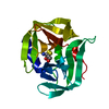
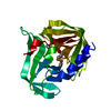
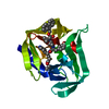
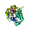
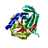
| Entry | Database: PDB / ID: 2zu1 | ||||||
|---|---|---|---|---|---|---|---|
| Title | crystal structure of CVB3 3C protease mutant C147A | ||||||
 Components Components | 3C proteinase | ||||||
 Keywords Keywords | HYDROLASE / protease / Thiol protease | ||||||
| Function / homology |  Function and homology information Function and homology informationsymbiont-mediated perturbation of host transcription / symbiont-mediated suppression of host cytoplasmic pattern recognition receptor signaling pathway via inhibition of RIG-I activity / symbiont-mediated suppression of host cytoplasmic pattern recognition receptor signaling pathway via inhibition of MDA-5 activity / symbiont-mediated suppression of host cytoplasmic pattern recognition receptor signaling pathway via inhibition of MAVS activity / picornain 2A / symbiont-mediated suppression of host mRNA export from nucleus / symbiont genome entry into host cell via pore formation in plasma membrane / picornain 3C / T=pseudo3 icosahedral viral capsid / host cell cytoplasmic vesicle membrane ...symbiont-mediated perturbation of host transcription / symbiont-mediated suppression of host cytoplasmic pattern recognition receptor signaling pathway via inhibition of RIG-I activity / symbiont-mediated suppression of host cytoplasmic pattern recognition receptor signaling pathway via inhibition of MDA-5 activity / symbiont-mediated suppression of host cytoplasmic pattern recognition receptor signaling pathway via inhibition of MAVS activity / picornain 2A / symbiont-mediated suppression of host mRNA export from nucleus / symbiont genome entry into host cell via pore formation in plasma membrane / picornain 3C / T=pseudo3 icosahedral viral capsid / host cell cytoplasmic vesicle membrane / ribonucleoside triphosphate phosphatase activity / nucleoside-triphosphate phosphatase / channel activity / monoatomic ion transmembrane transport / symbiont-mediated suppression of host NF-kappaB cascade / host cell cytoplasm / DNA replication / RNA helicase activity / endocytosis involved in viral entry into host cell / symbiont-mediated activation of host autophagy / RNA-directed RNA polymerase / cysteine-type endopeptidase activity / viral RNA genome replication / nucleotide binding / RNA-directed RNA polymerase activity / symbiont entry into host cell / virion attachment to host cell / host cell nucleus / DNA-templated transcription / structural molecule activity / proteolysis / RNA binding / zinc ion binding / ATP binding Similarity search - Function | ||||||
| Biological species |   Human coxsackievirus B3 Human coxsackievirus B3 | ||||||
| Method |  X-RAY DIFFRACTION / SYNCHROTRON / MOLECULAR REPLACEMENT / Resolution: 1.38 Å X-RAY DIFFRACTION / SYNCHROTRON / MOLECULAR REPLACEMENT / Resolution: 1.38 Å | ||||||
 Authors Authors | Lee, C.C. / Tsui, Y.C. / Wang, A.H.-J. | ||||||
 Citation Citation | Journal: J.Biol.Chem. / Year: 2009 Title: Structural Basis of Inhibition Specificities of 3C and 3C-like Proteases by Zinc-coordinating and Peptidomimetic Compounds Authors: Lee, C.C. / Kuo, C.J. / Ko, T.P. / Hsu, M.F. / Tsui, Y.C. / Chang, S.C. / Yang, S. / Chen, S.J. / Chen, H.C. / Hsu, M.C. / Shih, S.R. / Liang, P.H. / Wang, A.H.-J. | ||||||
| History |
|
- Structure visualization
| Structure viewer | Molecule: MolmilJmol/JSmol |
|---|
- Downloads & links
-Download
| PDBx/mmCIF format | 2zu1.cif.gz | 95 KB | Display | PDBx/mmCIF format |
|---|---|---|---|---|
| PDB format | pdb2zu1.ent.gz | 71.5 KB | Display | PDB format |
| PDBx/mmJSON format | 2zu1.json.gz | Tree view | PDBx/mmJSON format | |
| Others |  Other downloads Other downloads |
-Validation report
| Arichive directory | https://data.pdbj.org/pub/pdb/validation_reports/zu/2zu1ftp://data.pdbj.org/pub/pdb/validation_reports/zu/2zu1 | HTTPS FTP |
|---|
-Related structure data
| Related structure data | 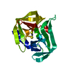 2ztxSC 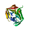 2ztyC 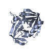 2ztzC 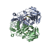 2zu2C 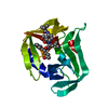 2zu3C 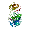 2zu4C 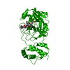 2zu5C S: Starting model for refinement C: citing same article ( |
|---|---|
| Similar structure data |


-Links
 PDBj
PDBj

- Assembly
| Deposited unit | 
| ||||||||
|---|---|---|---|---|---|---|---|---|---|
| 1 | 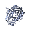
| ||||||||
| 2 | 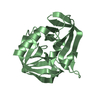
| ||||||||
| 3 |
| ||||||||
| Unit cell |
|
-Components
| #1: Protein | Mass: 20266.229 Da / Num. of mol.: 2 / Mutation: C147A Source method: isolated from a genetically manipulated source Source: (gene. exp.) Human coxsackievirus B3 / Plasmid: pET16b / Production host:  References: UniProt: Q90092, UniProt: P03313*PLUS, picornain 3C #2: Water | ChemComp-HOH / |  Mass: 18.015 Da / Num. of mol.: 624 / Source method: isolated from a natural source / Formula: H2O Mass: 18.015 Da / Num. of mol.: 624 / Source method: isolated from a natural source / Formula: H2O |
|---|
-Experimental details
-Experiment
| Experiment | Method: X-RAY DIFFRACTION / Number of used crystals: 1 |
|---|
- Sample preparation
| Crystal | Density Matthews: 2.14 Å3/Da / Density % sol: 42.44 % |
|---|---|
| Crystal grow | Temperature: 286 K / Method: vapor diffusion, sitting drop / pH: 8 Details: 24~30% PEG 4000, 0.2M magnesium chloride, 0.1M Tris-HCl, pH 8.0, VAPOR DIFFUSION, SITTING DROP, temperature 286 K |
-Data collection
| Diffraction | Mean temperature: 100 K |
|---|---|
| Diffraction source | Source: SYNCHROTRON / Site: NSRRC  / Beamline: BL13B1 / Beamline: BL13B1 |
| Detector | Type: ADSC QUANTUM 315 / Detector: CCD / Date: Sep 20, 2006 |
| Radiation | Protocol: SINGLE WAVELENGTH / Monochromatic (M) / Laue (L): M / Scattering type: x-ray |
| Radiation wavelength | Relative weight: 1 |
| Reflection | Resolution: 1.38→30 Å / Num. all: 69749 / Num. obs: 69121 / % possible obs: 99.1 % / Observed criterion σ(I): 1 / Redundancy: 3.3 % / Rmerge(I) obs: 0.063 / Net I/σ(I): 19.9 |
| Reflection shell | Resolution: 1.38→1.43 Å / Redundancy: 2.7 % / Rmerge(I) obs: 0.578 / Mean I/σ(I) obs: 2.1 / Num. unique all: 6740 / % possible all: 96.8 |
- Processing
| Software |
| |||||||||||||||||||||||||
|---|---|---|---|---|---|---|---|---|---|---|---|---|---|---|---|---|---|---|---|---|---|---|---|---|---|---|
| Refinement | Method to determine structure: MOLECULAR REPLACEMENT Starting model: PDB ENTRY 2ZTX Resolution: 1.38→30 Å / Occupancy max: 1 / Occupancy min: 1 / Isotropic thermal model: Overall / Cross valid method: THROUGHOUT / σ(F): 0 / σ(I): 0
| |||||||||||||||||||||||||
| Solvent computation | Bsol: 69.309 Å2 | |||||||||||||||||||||||||
| Displacement parameters | Biso max: 53.47 Å2 / Biso mean: 18.2 Å2 / Biso min: 7.39 Å2
| |||||||||||||||||||||||||
| Refinement step | Cycle: LAST / Resolution: 1.38→30 Å
| |||||||||||||||||||||||||
| Refine LS restraints |
|
