| Entry | Database: PDB / ID: 4ydr
|
|---|
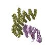
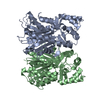
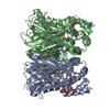
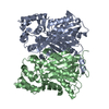
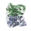
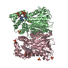
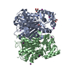
| Title | Crystal structure of oxidized homoserine dehydrogenase of Sulfolobus tokodaii |
|---|
 Components Components | Homoserine dehydrogenase |
|---|
 Keywords Keywords | OXIDOREDUCTASE / Oxidized form |
|---|
| Function / homology |  Function and homology information Function and homology information
homoserine dehydrogenase / homoserine dehydrogenase activity / L-threonine biosynthetic process / : / NADP binding / metal ion bindingSimilarity search - Function Homoserine dehydrogenase lacking ACT domain / Homoserine dehydrogenase, conserved site / Homoserine dehydrogenase signature. / Homoserine dehydrogenase, catalytic / Homoserine dehydrogenase / Aspartate/homoserine dehydrogenase, NAD-binding / Homoserine dehydrogenase, NAD binding domain / Dihydrodipicolinate Reductase; domain 2 / Dihydrodipicolinate Reductase; domain 2 / NAD(P)-binding Rossmann-like Domain ...Homoserine dehydrogenase lacking ACT domain / Homoserine dehydrogenase, conserved site / Homoserine dehydrogenase signature. / Homoserine dehydrogenase, catalytic / Homoserine dehydrogenase / Aspartate/homoserine dehydrogenase, NAD-binding / Homoserine dehydrogenase, NAD binding domain / Dihydrodipicolinate Reductase; domain 2 / Dihydrodipicolinate Reductase; domain 2 / NAD(P)-binding Rossmann-like Domain / NAD(P)-binding domain superfamily / Rossmann fold / 2-Layer Sandwich / 3-Layer(aba) Sandwich / Alpha BetaSimilarity search - Domain/homology |
|---|
| Biological species |   Sulfolobus tokodaii (archaea) Sulfolobus tokodaii (archaea) |
|---|
| Method |  X-RAY DIFFRACTION / SYNCHROTRON / MOLECULAR REPLACEMENT / Resolution: 1.6 Å X-RAY DIFFRACTION / SYNCHROTRON / MOLECULAR REPLACEMENT / Resolution: 1.6 Å |
|---|
 Authors Authors | Goto, M. / Yoshimune, K. / Kaneko, R. |
|---|
 Citation Citation | Journal: Biochem Biophys Rep / Year: 2015
Title: Structural insight into activation of homoserine dehydrogenase from the archaeonSulfolobus tokodaiivia reduction.
Authors: Tomonaga, Y. / Kaneko, R. / Goto, M. / Ohshima, T. / Yoshimune, K. |
|---|
| History | | Deposition | Feb 23, 2015 | Deposition site: RCSB / Processing site: PDBJ |
|---|
| Revision 1.0 | Nov 11, 2015 | Provider: repository / Type: Initial release |
|---|
| Revision 1.1 | Dec 18, 2019 | Group: Data collection / Database references / Derived calculations
Category: citation / diffrn_source / pdbx_struct_oper_list
Item: _citation.pdbx_database_id_PubMed / _citation.title ..._citation.pdbx_database_id_PubMed / _citation.title / _diffrn_source.pdbx_synchrotron_site / _pdbx_struct_oper_list.symmetry_operation |
|---|
| Revision 1.2 | Oct 30, 2024 | Group: Data collection / Database references ...Data collection / Database references / Derived calculations / Structure summary
Category: chem_comp_atom / chem_comp_bond ...chem_comp_atom / chem_comp_bond / database_2 / pdbx_entry_details / pdbx_modification_feature / pdbx_struct_conn_angle / struct_conn
Item: _database_2.pdbx_DOI / _database_2.pdbx_database_accession ..._database_2.pdbx_DOI / _database_2.pdbx_database_accession / _pdbx_struct_conn_angle.ptnr1_auth_comp_id / _pdbx_struct_conn_angle.ptnr1_auth_seq_id / _pdbx_struct_conn_angle.ptnr1_label_atom_id / _pdbx_struct_conn_angle.ptnr1_label_comp_id / _pdbx_struct_conn_angle.ptnr1_label_seq_id / _pdbx_struct_conn_angle.ptnr2_symmetry / _pdbx_struct_conn_angle.ptnr3_auth_comp_id / _pdbx_struct_conn_angle.ptnr3_auth_seq_id / _pdbx_struct_conn_angle.ptnr3_label_atom_id / _pdbx_struct_conn_angle.ptnr3_label_comp_id / _pdbx_struct_conn_angle.ptnr3_label_seq_id / _pdbx_struct_conn_angle.value / _struct_conn.pdbx_dist_value / _struct_conn.ptnr1_auth_comp_id / _struct_conn.ptnr1_auth_seq_id / _struct_conn.ptnr1_label_atom_id / _struct_conn.ptnr1_label_comp_id / _struct_conn.ptnr1_label_seq_id / _struct_conn.ptnr2_symmetry |
|---|
|
|---|
 Movie
Movie Controller

 Yorodumi
 Open data
 Basic information
 Structure visualization
 Downloads & links
 Other downloads
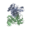


 PDBj
PDBj Assembly
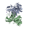


 Mass: 22.990 Da / Num. of mol.: 1 / Source method: obtained synthetically / Formula: Na
 Mass: 18.015 Da / Num. of mol.: 402 / Source method: isolated from a natural source / Formula: H2O
 Sample preparation
 / Beamline: AR-NE3A / Wavelength: 1 Å
 Processing