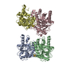
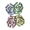
Protein / Protein/peptide , 2 types, 4 molecules ABCD
#1: Protein
Bifunctionallysine-specificdemethylaseandhistidyl-hydroxylaseNO66 / 60S ribosomal protein L8 histidine hydroxylase / Histone lysine demethylase NO66 / Myc-associated ...60S ribosomal protein L8 histidine hydroxylase / Histone lysine demethylase NO66 / Myc-associated protein with JmjC domain / Nucleolar protein 66 / hsNO66 / Ribosomal oxygenase NO66 / ROX
Mass: 52984.836 Da / Num. of mol.: 2 / Fragment: UNP RESIDUES 176-641 Source method: isolated from a genetically manipulated source Source: (gene. exp.) Homo sapiens (human) / Gene: NO66, C14orf169, MAPJD / Production host: Escherichia coli (E. coli) References: UniProt: Q9H6W3, Oxidoreductases; Acting on paired donors, with incorporation or reduction of molecular oxygen; With 2-oxoglutarate as one donor, and incorporation of one atom of oxygen ...References: UniProt: Q9H6W3, Oxidoreductases; Acting on paired donors, with incorporation or reduction of molecular oxygen; With 2-oxoglutarate as one donor, and incorporation of one atom of oxygen into each donor, [histone H3]-dimethyl-L-lysine36 demethylase
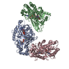
#2: Protein/peptide
Rpl8peptide
Mass: 1104.200 Da / Num. of mol.: 2 / Source method: obtained synthetically / Source: (synth.) Homo sapiens (human) / References: UniProt: P62917*PLUS
Protocol: SINGLE WAVELENGTH / Monochromatic (M) / Laue (L): M / Scattering type: x-ray
Radiation wavelength
Wavelength: 0.9791 Å / Relative weight: 1
Reflection
Resolution: 2.2→50 Å / Num. obs: 62029 / % possible obs: 98 % / Redundancy: 3.4 % / Rmerge(I) obs: 0.123 / Net I/σ(I): 7.6
Reflection shell
Resolution: 2.2→2.32 Å / Redundancy: 3.4 % / Rmerge(I) obs: 0.502 / Mean I/σ(I) obs: 2.4 / % possible all: 97.8
-
Processing
Software
Name
Version
Classification
REFMAC
5.7.0032
refinement
MOSFLM
dataprocessing
SCALA
datascaling
PHASER
phasing
Coot
modelbuilding
Refinement
Resolution: 2.2→40.57 Å / Cor.coef. Fo:Fc: 0.93 / Cor.coef. Fo:Fc free: 0.882 / SU B: 5.743 / SU ML: 0.146 / Cross valid method: THROUGHOUT / ESU R: 0.24 / ESU R Free: 0.213 / Stereochemistry target values: MAXIMUM LIKELIHOOD / Details: HYDROGENS HAVE BEEN ADDED IN THE RIDING POSITIONS
Rfactor
Num. reflection
% reflection
Selection details
Rfree
0.26154
3314
5.1 %
RANDOM
Rwork
0.20279
-
-
-
obs
0.20578
62029
97.86 %
-
Solvent computation
Ion probe radii: 0.8 Å / Shrinkage radii: 0.8 Å / VDW probe radii: 1.2 Å / Solvent model: MASK
 Movie
Movie Controller

 Yorodumi
 Open data
 Basic information
 Components
 Keywords
 Function and homology information
 Homo sapiens (human)
 X-RAY DIFFRACTION /
  Authors
 Citation
 Structure visualization
 Downloads & links
 Other downloads
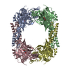


 PDBj
PDBj


 Assembly
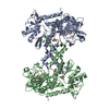


 Mass: 58.693 Da / Num. of mol.: 2 / Source method: obtained synthetically / Formula: Ni
 Mass: 147.086 Da / Num. of mol.: 2 / Source method: obtained synthetically / Formula: C4H5NO5 / Comment: inhibitor*YM
 Mass: 92.094 Da / Num. of mol.: 3 / Source method: obtained synthetically / Formula: C3H8O3
 Mass: 59.044 Da / Num. of mol.: 1 / Source method: obtained synthetically / Formula: C2H3O2
 Sample preparation
 / Beamline: BL17U / Wavelength: 0.9791 Å
 Processing