 Movie
Movie Controller

[English] 日本語
 Yorodumi
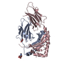
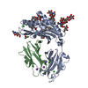
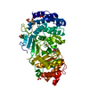
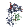
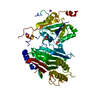
- PDB-4y2o: Structure of CFA/I pili chaperone-major subunit complex (CfaA-CfaB) -
+ Open data

- Basic information
| Entry | Database: PDB / ID: 4y2o | ||||||
|---|---|---|---|---|---|---|---|
| Title | Structure of CFA/I pili chaperone-major subunit complex (CfaA-CfaB) | ||||||
 Components Components |
| ||||||
 Keywords Keywords | STRUCTURAL PROTEIN / Enterotoxigenic Escherichia coli / periplasmic chaperone / major pilin / self-assembly / fimbriae | ||||||
| Function / homology |  Function and homology information Function and homology information | ||||||
| Biological species | Escherichia coli O78:H11 | ||||||
| Method |  X-RAY DIFFRACTION / SYNCHROTRON / MOLECULAR REPLACEMENT / Resolution: 2.419 Å X-RAY DIFFRACTION / SYNCHROTRON / MOLECULAR REPLACEMENT / Resolution: 2.419 Å | ||||||
 Authors Authors | Bao, R. / Xia, D. | ||||||
 Citation Citation | Journal: Mol. Microbiol. / Year: 2016 Title: Off-pathway assembly of fimbria subunits is prevented by chaperone CfaA of CFA/I fimbriae from enterotoxigenic E. coli. Authors: Bao, R. / Liu, Y. / Savarino, S.J. / Xia, D. | ||||||
| History |
|
- Structure visualization
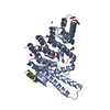
| Structure viewer | Molecule: MolmilJmol/JSmol |
|---|
- Downloads & links
-Download
| PDBx/mmCIF format | 4y2o.cif.gz | 89.9 KB | Display | PDBx/mmCIF format |
|---|---|---|---|---|
| PDB format | pdb4y2o.ent.gz | 66.2 KB | Display | PDB format |
| PDBx/mmJSON format | 4y2o.json.gz | Tree view | PDBx/mmJSON format | |
| Others |  Other downloads Other downloads |
-Validation report
| Arichive directory | https://data.pdbj.org/pub/pdb/validation_reports/y2/4y2oftp://data.pdbj.org/pub/pdb/validation_reports/y2/4y2o | HTTPS FTP |
|---|
-Related structure data
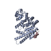
| Related structure data |  4y2lC  4y2nC  3f84S C: citing same article ( S: Starting model for refinement |
|---|---|
| Similar structure data |


-Links
 PDBj
PDBj- Assembly
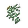
| Deposited unit | 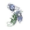
| ||||||||
|---|---|---|---|---|---|---|---|---|---|
| 1 |
| ||||||||
| Unit cell |
| ||||||||
| Components on special symmetry positions |
|
-Components
| #1: Protein | Mass: 26493.488 Da / Num. of mol.: 1 / Fragment: UNP residues 19-238 / Mutation: T112I Source method: isolated from a genetically manipulated source Source: (gene. exp.)  Strain: H10407 / ETEC / Gene: cfaA, ETEC_p948_0390 / Production host: | ||||||
|---|---|---|---|---|---|---|---|
| #2: Protein | Mass: 14715.560 Da / Num. of mol.: 1 / Fragment: UNP residues 38-170 Source method: isolated from a genetically manipulated source Source: (gene. exp.) Strain: H10407 / ETEC / Gene: cfaB, ETEC_p948_0400 / Production host: | ||||||
| #3: Chemical |   Mass: 150.173 Da / Num. of mol.: 2 / Source method: obtained synthetically / Formula: C6H14O4 Mass: 150.173 Da / Num. of mol.: 2 / Source method: obtained synthetically / Formula: C6H14O4#4: Chemical |   Mass: 58.693 Da / Num. of mol.: 2 / Source method: obtained synthetically / Formula: Ni Mass: 58.693 Da / Num. of mol.: 2 / Source method: obtained synthetically / Formula: Ni#5: Water | ChemComp-HOH / |  Mass: 18.015 Da / Num. of mol.: 139 / Source method: isolated from a natural source / Formula: H2O Mass: 18.015 Da / Num. of mol.: 139 / Source method: isolated from a natural source / Formula: H2OHas protein modification | Y | |
-Experimental details
-Experiment
| Experiment | Method: X-RAY DIFFRACTION / Number of used crystals: 1 |
|---|
- Sample preparation
| Crystal | Density Matthews: 2.93 Å3/Da / Density % sol: 58.03 % |
|---|---|
| Crystal grow | Temperature: 293 K / Method: vapor diffusion, hanging drop / Details: Na2HPO4-NaH2PO4 pH 6.6, 9% PEG8000 |
-Data collection
| Diffraction | Mean temperature: 100 K |
|---|---|
| Diffraction source | Source: SYNCHROTRON / Site: APS  / Beamline: 22-BM / Wavelength: 1 Å / Beamline: 22-BM / Wavelength: 1 Å |
| Detector | Type: MARMOSAIC 225 mm CCD / Detector: CCD / Date: Jun 15, 2012 |
| Radiation | Protocol: SINGLE WAVELENGTH / Monochromatic (M) / Laue (L): M / Scattering type: x-ray |
| Radiation wavelength | Wavelength: 1 Å / Relative weight: 1 |
| Reflection | Resolution: 2.32→44.464 Å / Num. obs: 18960 / % possible obs: 96.4 % / Redundancy: 7.5 % / Rmerge(I) obs: 0.079 / Net I/σ(I): 20 |
| Reflection shell | Resolution: 2.32→2.4 Å / Redundancy: 2.3 % / Rmerge(I) obs: 0.474 / Mean I/σ(I) obs: 1 / % possible all: 72.2 |
- Processing
| Software |
| |||||||||||||||||||||||||||||||||||||||||||||||||
|---|---|---|---|---|---|---|---|---|---|---|---|---|---|---|---|---|---|---|---|---|---|---|---|---|---|---|---|---|---|---|---|---|---|---|---|---|---|---|---|---|---|---|---|---|---|---|---|---|---|---|
| Refinement | Method to determine structure: MOLECULAR REPLACEMENT Starting model: 3F84 Resolution: 2.419→44.464 Å / Cross valid method: FREE R-VALUE / σ(F): 1.14 / Phase error: 31.2 / Stereochemistry target values: TWIN_LSQ_F
| |||||||||||||||||||||||||||||||||||||||||||||||||
| Solvent computation | Shrinkage radii: 0.9 Å / VDW probe radii: 1.11 Å / Solvent model: FLAT BULK SOLVENT MODEL | |||||||||||||||||||||||||||||||||||||||||||||||||
| Refinement step | Cycle: LAST / Resolution: 2.419→44.464 Å
| |||||||||||||||||||||||||||||||||||||||||||||||||
| Refine LS restraints |
| |||||||||||||||||||||||||||||||||||||||||||||||||
| LS refinement shell |
|
