 Movie
Movie Controller

[English] 日本語
 Yorodumi
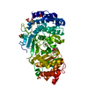
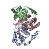
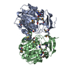
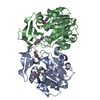
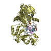
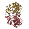
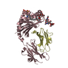
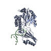
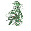
- PDB-5h3l: Structure of methylglyoxal synthase crystallised as a contaminant -
+ Open data

- Basic information
| Entry | Database: PDB / ID: 5h3l | ||||||
|---|---|---|---|---|---|---|---|
| Title | Structure of methylglyoxal synthase crystallised as a contaminant | ||||||
 Components Components | Methylglyoxal synthase | ||||||
 Keywords Keywords | LYASE / Contaminant | ||||||
| Function / homology | Methylglyoxal synthase-like domain / Rossmann fold / 3-Layer(aba) Sandwich / Alpha Beta / FORMIC ACID Function and homology information Function and homology information | ||||||
| Biological species | synthetic construct (others) | ||||||
| Method |  X-RAY DIFFRACTION / SYNCHROTRON / MOLECULAR REPLACEMENT / Resolution: 2.1 Å X-RAY DIFFRACTION / SYNCHROTRON / MOLECULAR REPLACEMENT / Resolution: 2.1 Å | ||||||
 Authors Authors | Hatti, K. / Dadireddy, V. / Srinivasan, N. / Ramakumar, S. / Murthy, M.R.N. | ||||||
 Citation Citation | Journal: J. Struct. Biol. / Year: 2017 Title: Structure determination of contaminant proteins using the MarathonMR procedure. Authors: Hatti, K. / Biswas, A. / Chaudhary, S. / Dadireddy, V. / Sekar, K. / Srinivasan, N. / Murthy, M.R. | ||||||
| History |
|
- Structure visualization
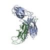
| Structure viewer | Molecule: MolmilJmol/JSmol |
|---|
- Downloads & links
-Download
| PDBx/mmCIF format | 5h3l.cif.gz | 99.1 KB | Display | PDBx/mmCIF format |
|---|---|---|---|---|
| PDB format | pdb5h3l.ent.gz | 75.8 KB | Display | PDB format |
| PDBx/mmJSON format | 5h3l.json.gz | Tree view | PDBx/mmJSON format | |
| Others |  Other downloads Other downloads |
-Validation report
| Arichive directory | https://data.pdbj.org/pub/pdb/validation_reports/h3/5h3lftp://data.pdbj.org/pub/pdb/validation_reports/h3/5h3l | HTTPS FTP |
|---|
-Related structure data
| Related structure data |  5h4fC  5h4gC  5h4hC  1b93S S: Starting model for refinement C: citing same article ( |
|---|---|
| Similar structure data |
-Links
 PDBj
PDBj- Assembly
| Deposited unit | 
| ||||||||
|---|---|---|---|---|---|---|---|---|---|
| 1 |
| ||||||||
| Unit cell |
|
-Components
| #1: Protein | Mass: 16958.518 Da / Num. of mol.: 3 Source method: isolated from a genetically manipulated source Source: (gene. exp.) synthetic construct (others) / Production host:  #2: Chemical |   Mass: 46.025 Da / Num. of mol.: 3 / Source method: obtained synthetically / Formula: CH2O2 Mass: 46.025 Da / Num. of mol.: 3 / Source method: obtained synthetically / Formula: CH2O2#3: Water | ChemComp-HOH / |  Mass: 18.015 Da / Num. of mol.: 125 / Source method: isolated from a natural source / Formula: H2O Mass: 18.015 Da / Num. of mol.: 125 / Source method: isolated from a natural source / Formula: H2OHas protein modification | Y | |
|---|
-Experimental details
-Experiment
| Experiment | Method: X-RAY DIFFRACTION / Number of used crystals: 1 |
|---|
- Sample preparation
| Crystal | Density Matthews: 2.43 Å3/Da / Density % sol: 49.35 % |
|---|---|
| Crystal grow | Temperature: 295 K / Method: vapor diffusion, sitting drop / pH: 7 / Details: 3.5M Sodium formate.HCl, pH 7.0 |
-Data collection
| Diffraction | Mean temperature: 100 K |
|---|---|
| Diffraction source | Source: SYNCHROTRON / Site: ESRF  / Beamline: BM14 / Wavelength: 0.95372 Å / Beamline: BM14 / Wavelength: 0.95372 Å |
| Detector | Type: MARMOSAIC 225 mm CCD / Detector: CCD / Date: Jul 8, 2016 |
| Radiation | Protocol: SINGLE WAVELENGTH / Monochromatic (M) / Laue (L): M / Scattering type: x-ray |
| Radiation wavelength | Wavelength: 0.95372 Å / Relative weight: 1 |
| Reflection | Resolution: 2.1→61.16 Å / Num. all: 30046 / Num. obs: 29980 / % possible obs: 100 % / Redundancy: 14.4 % / CC1/2: 0.991 / Net I/σ(I): 8 |
| Reflection shell | Resolution: 2.1→2.175 Å / Redundancy: 14.6 % / Rmerge(I) obs: 2.179 / Mean I/σ(I) obs: 1.4 / CC1/2: 0.436 / % possible all: 100 |
- Processing
| Software |
| ||||||||||||||||||||||||||||||||||||||||||||||||||||||||||||||||||||||||||||||||||||
|---|---|---|---|---|---|---|---|---|---|---|---|---|---|---|---|---|---|---|---|---|---|---|---|---|---|---|---|---|---|---|---|---|---|---|---|---|---|---|---|---|---|---|---|---|---|---|---|---|---|---|---|---|---|---|---|---|---|---|---|---|---|---|---|---|---|---|---|---|---|---|---|---|---|---|---|---|---|---|---|---|---|---|---|---|---|
| Refinement | Method to determine structure: MOLECULAR REPLACEMENT Starting model: 1B93 Resolution: 2.1→61.165 Å / SU ML: 0.3 / Cross valid method: FREE R-VALUE / σ(F): 1.34 / Phase error: 23.35
| ||||||||||||||||||||||||||||||||||||||||||||||||||||||||||||||||||||||||||||||||||||
| Solvent computation | Shrinkage radii: 0.9 Å / VDW probe radii: 1.11 Å | ||||||||||||||||||||||||||||||||||||||||||||||||||||||||||||||||||||||||||||||||||||
| Refinement step | Cycle: LAST / Resolution: 2.1→61.165 Å
| ||||||||||||||||||||||||||||||||||||||||||||||||||||||||||||||||||||||||||||||||||||
| Refine LS restraints |
| ||||||||||||||||||||||||||||||||||||||||||||||||||||||||||||||||||||||||||||||||||||
| LS refinement shell |
|
