 Movie
Movie Controller

[English] 日本語
 Yorodumi
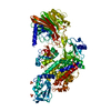
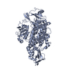
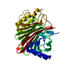
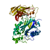
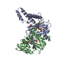
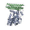
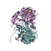
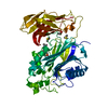
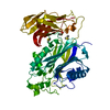
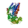
- PDB-6yhn: Crystal structure of domains 4-5 of CNFy from Yersinia pseudotube... -
+ Open data

- Basic information
| Entry | Database: PDB / ID: 6yhn | ||||||
|---|---|---|---|---|---|---|---|
| Title | Crystal structure of domains 4-5 of CNFy from Yersinia pseudotuberculosis | ||||||
 Components Components | Cytotoxic necrotizing factor | ||||||
 Keywords Keywords | TOXIN / CNF / cytotoxic necrotizing factor / deamidase / RhoA modification / RhoA activation / putative ADP-ribosyltransferase | ||||||
| Function / homology |  Function and homology information Function and homology information | ||||||
| Biological species |  Yersinia pseudotuberculosis (bacteria) Yersinia pseudotuberculosis (bacteria) | ||||||
| Method |  X-RAY DIFFRACTION / SYNCHROTRON / MOLECULAR REPLACEMENT / molecular replacement / Resolution: 1.8 Å X-RAY DIFFRACTION / SYNCHROTRON / MOLECULAR REPLACEMENT / molecular replacement / Resolution: 1.8 Å | ||||||
| Model details | Structure comprises residues 1-1014, active site cysteine 866 has been mutated to serine | ||||||
 Authors Authors | Lukat, P. / Gazdag, E.M. / Heidler, T.V. / Blankenfeldt, W. | ||||||
 Citation Citation | Journal: Embo J. / Year: 2021 Title: Crystal structure of bacterial cytotoxic necrotizing factor CNF Y reveals molecular building blocks for intoxication. Authors: Chaoprasid, P. / Lukat, P. / Muhlen, S. / Heidler, T. / Gazdag, E.M. / Dong, S. / Bi, W. / Ruter, C. / Kirchenwitz, M. / Steffen, A. / Jansch, L. / Stradal, T.E.B. / Dersch, P. / Blankenfeldt, W. | ||||||
| History |
|
- Structure visualization
| Structure viewer | Molecule: MolmilJmol/JSmol |
|---|
- Downloads & links
-Download
| PDBx/mmCIF format | 6yhn.cif.gz | 289.2 KB | Display | PDBx/mmCIF format |
|---|---|---|---|---|
| PDB format | pdb6yhn.ent.gz | 233.7 KB | Display | PDB format |
| PDBx/mmJSON format | 6yhn.json.gz | Tree view | PDBx/mmJSON format | |
| Others |  Other downloads Other downloads |
-Validation report
| Arichive directory | https://data.pdbj.org/pub/pdb/validation_reports/yh/6yhnftp://data.pdbj.org/pub/pdb/validation_reports/yh/6yhn | HTTPS FTP |
|---|
-Related structure data

-Links
 PDBj
PDBj- Assembly
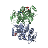
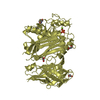
| Deposited unit | 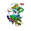
| ||||||||
|---|---|---|---|---|---|---|---|---|---|
| 1 |
| ||||||||
| Unit cell |
|
-Components
-Protein , 1 types, 1 molecules A
| #1: Protein | Mass: 57848.938 Da / Num. of mol.: 1 / Fragment: CNFy residues 526-1014 Source method: isolated from a genetically manipulated source Details: Fragment of CNFy comprising residues 526-1014. Cys 866 has found to be oxidized in the structure. Source: (gene. exp.) Yersinia pseudotuberculosis (bacteria) / Gene: cnf / Plasmid: pCOLA-Duet-1Details (production host): pCOLA-Duet-1 has been modified with sequence encoding for N-terminal Strep-tag II and TEV protease cleavage site and C-terminal 3 x FLAG epitope Production host: |
|---|
-Non-polymers , 6 types, 376 molecules 


| #2: Chemical | ChemComp-BU3 / ( Mass: 90.121 Da / Num. of mol.: 1 / Source method: obtained synthetically / Formula: C4H10O2 Mass: 90.121 Da / Num. of mol.: 1 / Source method: obtained synthetically / Formula: C4H10O2 | ||||||||
|---|---|---|---|---|---|---|---|---|---|
| #3: Chemical | ChemComp-MG /  Mass: 24.305 Da / Num. of mol.: 4 / Source method: obtained synthetically / Formula: Mg Mass: 24.305 Da / Num. of mol.: 4 / Source method: obtained synthetically / Formula: Mg#4: Chemical |  Mass: 22.990 Da / Num. of mol.: 2 / Source method: obtained synthetically / Formula: Na Mass: 22.990 Da / Num. of mol.: 2 / Source method: obtained synthetically / Formula: Na#5: Chemical | ChemComp-CL / |  Mass: 35.453 Da / Num. of mol.: 1 / Source method: obtained synthetically / Formula: Cl Mass: 35.453 Da / Num. of mol.: 1 / Source method: obtained synthetically / Formula: Cl#6: Chemical | ChemComp-SO4 / |  Mass: 96.063 Da / Num. of mol.: 1 / Source method: obtained synthetically / Formula: SO4 Mass: 96.063 Da / Num. of mol.: 1 / Source method: obtained synthetically / Formula: SO4#7: Water | ChemComp-HOH / | Mass: 18.015 Da / Num. of mol.: 367 / Source method: isolated from a natural source / Formula: H2O |
-Details
| Has ligand of interest | N |
|---|---|
| Has protein modification | Y |
-Experimental details
-Experiment
| Experiment | Method: X-RAY DIFFRACTION / Number of used crystals: 1 |
|---|
- Sample preparation
| Crystal | Density Matthews: 2.19 Å3/Da / Density % sol: 43.88 % / Mosaicity: 0.1 ° |
|---|---|
| Crystal grow | Temperature: 293 K / Method: vapor diffusion Details: 0.24 M magnesium chloride, 22.5 % (w/v) PEG 2000 MME, 1 mM ATP |
-Data collection
| Diffraction | Mean temperature: 100 K / Serial crystal experiment: N | ||||||||||||||||||||||||
|---|---|---|---|---|---|---|---|---|---|---|---|---|---|---|---|---|---|---|---|---|---|---|---|---|---|
| Diffraction source | Source: SYNCHROTRON / Site: SLS  / Beamline: X06DA / Wavelength: 1 Å / Beamline: X06DA / Wavelength: 1 Å | ||||||||||||||||||||||||
| Detector | Type: DECTRIS PILATUS 2M-F / Detector: PIXEL / Date: Apr 12, 2018 | ||||||||||||||||||||||||
| Radiation | Protocol: SINGLE WAVELENGTH / Monochromatic (M) / Laue (L): M / Scattering type: x-ray | ||||||||||||||||||||||||
| Radiation wavelength | Wavelength: 1 Å / Relative weight: 1 | ||||||||||||||||||||||||
| Reflection | Resolution: 1.8→48.27 Å / Num. obs: 49960 / % possible obs: 99.9 % / Redundancy: 32.7 % / CC1/2: 0.982 / Rmerge(I) obs: 0.257 / Rpim(I) all: 0.046 / Rrim(I) all: 0.261 / Net I/σ(I): 12.4 | ||||||||||||||||||||||||
| Reflection shell | Diffraction-ID: 1
|
-Phasing
| Phasing | Method: molecular replacement |
|---|
- Processing
| Software |
| |||||||||||||||||||||||||||||||||||||||||||||||||||||||||||||||||||||||||||||||||||||||||||||||||||||||||||||||||||||||||||||||||||||||||||||||||||||||||||||||||||||||||||||||
|---|---|---|---|---|---|---|---|---|---|---|---|---|---|---|---|---|---|---|---|---|---|---|---|---|---|---|---|---|---|---|---|---|---|---|---|---|---|---|---|---|---|---|---|---|---|---|---|---|---|---|---|---|---|---|---|---|---|---|---|---|---|---|---|---|---|---|---|---|---|---|---|---|---|---|---|---|---|---|---|---|---|---|---|---|---|---|---|---|---|---|---|---|---|---|---|---|---|---|---|---|---|---|---|---|---|---|---|---|---|---|---|---|---|---|---|---|---|---|---|---|---|---|---|---|---|---|---|---|---|---|---|---|---|---|---|---|---|---|---|---|---|---|---|---|---|---|---|---|---|---|---|---|---|---|---|---|---|---|---|---|---|---|---|---|---|---|---|---|---|---|---|---|---|---|---|---|
| Refinement | Method to determine structure: MOLECULAR REPLACEMENT Starting model: Corresponding fragment from the full-length toxin Resolution: 1.8→45.265 Å / SU ML: 0.2 / Cross valid method: THROUGHOUT / σ(F): 1.34 / Phase error: 21.23
| |||||||||||||||||||||||||||||||||||||||||||||||||||||||||||||||||||||||||||||||||||||||||||||||||||||||||||||||||||||||||||||||||||||||||||||||||||||||||||||||||||||||||||||||
| Solvent computation | Shrinkage radii: 0.9 Å / VDW probe radii: 1.11 Å | |||||||||||||||||||||||||||||||||||||||||||||||||||||||||||||||||||||||||||||||||||||||||||||||||||||||||||||||||||||||||||||||||||||||||||||||||||||||||||||||||||||||||||||||
| Displacement parameters | Biso max: 150.78 Å2 / Biso mean: 38.201 Å2 / Biso min: 12.4 Å2 | |||||||||||||||||||||||||||||||||||||||||||||||||||||||||||||||||||||||||||||||||||||||||||||||||||||||||||||||||||||||||||||||||||||||||||||||||||||||||||||||||||||||||||||||
| Refinement step | Cycle: final / Resolution: 1.8→45.265 Å
| |||||||||||||||||||||||||||||||||||||||||||||||||||||||||||||||||||||||||||||||||||||||||||||||||||||||||||||||||||||||||||||||||||||||||||||||||||||||||||||||||||||||||||||||
| LS refinement shell | Refine-ID: X-RAY DIFFRACTION / Rfactor Rfree error: 0 / % reflection obs: 100 %
| |||||||||||||||||||||||||||||||||||||||||||||||||||||||||||||||||||||||||||||||||||||||||||||||||||||||||||||||||||||||||||||||||||||||||||||||||||||||||||||||||||||||||||||||
| Refinement TLS params. | Method: refined / Refine-ID: X-RAY DIFFRACTION
| |||||||||||||||||||||||||||||||||||||||||||||||||||||||||||||||||||||||||||||||||||||||||||||||||||||||||||||||||||||||||||||||||||||||||||||||||||||||||||||||||||||||||||||||
| Refinement TLS group |
|
