[English] 日本語
Yorodumi
- PDB-4wzw: Crystal structure of human Puf-A in complex with DNA -

+
Open data


ID or keywords:

Loading...

-
Basic information

Entry
Database: PDB / ID: 4wzw
TitleCrystal structure of human Puf-A in complex with DNA
Components
  • DNA (5'-D(P*CP*CP*CP*CP*CP*CP*CP*CP*CP*CP*CP*CP*CP*CP*CP*C)-3')
  • DNA (5'-D(P*GP*GP*GP*GP*GP*GP*GP*GP*GP*GP*GP*GP*GP*G)-3')
  • Pumilio domain-containing protein KIAA0020
KeywordsDNA Binding Protein / RNA Binding Protein/DNA / Pumilio repeat protein / RNA Binding Protein-DNA complex
Function / homology
Function and homology information


regulation of protein ADP-ribosylation / regulation of translation / chromosome / mRNA binding / nucleolus / endoplasmic reticulum / DNA binding / RNA binding / nucleoplasm
Similarity search - Function
CPL domain / Pumilio homologue 3 / CPL (NUC119) domain / Pumilio homology domain / Pumilio homology domain (PUM-HD) profile. / Pumilio RNA-binding repeat profile. / Pumilio RNA-binding repeat / Pumilio-like repeats / Armadillo-like helical / Armadillo-type fold
Similarity search - Domain/homology
DNA / DNA (> 10) / Pumilio homolog 3
Similarity search - Component
Biological speciesHomo sapiens (human)
synthetic construct (others)
MethodX-RAY DIFFRACTION / SYNCHROTRON / MOLECULAR REPLACEMENT / Resolution: 2.9516 Å
AuthorsQiu, C. / Hall, T.M.T.
Funding support United States, 1items
OrganizationGrant numberCountry
National Institutes of Health/National Institute of Environmental Health Sciences (NIH/NIEHS)Intramural Research Program United States
CitationJournal: Proc.Natl.Acad.Sci.USA / Year: 2014
Title: A divergent Pumilio repeat protein family for pre-rRNA processing and mRNA localization.
Authors: Qiu, C. / McCann, K.L. / Wine, R.N. / Baserga, S.J. / Hall, T.M.
History
DepositionNov 20, 2014Deposition site: RCSB / Processing site: RCSB
Revision 1.0Dec 31, 2014Provider: repository / Type: Initial release
Revision 1.1Jan 14, 2015Group: Database references
Revision 1.2Nov 27, 2019Group: Author supporting evidence / Database references ...Author supporting evidence / Database references / Derived calculations / Source and taxonomy
Category: citation / entity_src_gen ...citation / entity_src_gen / pdbx_audit_support / pdbx_entity_src_syn / pdbx_struct_oper_list
Item: _citation.journal_id_CSD / _entity_src_gen.pdbx_alt_source_flag ..._citation.journal_id_CSD / _entity_src_gen.pdbx_alt_source_flag / _pdbx_audit_support.funding_organization / _pdbx_entity_src_syn.pdbx_alt_source_flag / _pdbx_struct_oper_list.symmetry_operation
Revision 1.3Sep 27, 2023Group: Data collection / Database references / Refinement description
Category: chem_comp_atom / chem_comp_bond ...chem_comp_atom / chem_comp_bond / database_2 / pdbx_initial_refinement_model
Item: _database_2.pdbx_DOI / _database_2.pdbx_database_accession

-
Structure visualization

Structure viewerMolecule:
MolmilJmol/JSmol

Downloads & links

-
Assembly

Deposited unit
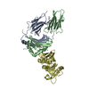
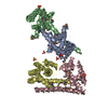
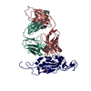
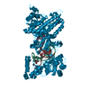
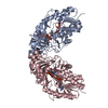
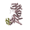
A: Pumilio domain-containing protein KIAA0020
B: DNA (5'-D(P*GP*GP*GP*GP*GP*GP*GP*GP*GP*GP*GP*GP*GP*G)-3')
C: DNA (5'-D(P*CP*CP*CP*CP*CP*CP*CP*CP*CP*CP*CP*CP*CP*CP*CP*C)-3')


Theoretical massNumber of molelcules
Total (without water)68,1963
Polymers68,1963
Non-polymers00
Water00
1


  • Idetical with deposited unit
  • defined by author&software
TypeNameSymmetry operationNumber
identity operation1_555x,y,z1
Buried area2580 Å2
ΔGint-23 kcal/mol
Surface area28740 Å2
MethodPISA
Unit cell
Length a, b, c (Å)69.122, 69.122, 273.831
Angle α, β, γ (deg.)90.00, 90.00, 90.00
Int Tables number95
Space group name H-MP4322

-
Components

#1: Protein Pumilio domain-containing protein KIAA0020 / HBV X-transactivated gene 5 protein / HBV XAg-transactivated protein 5 / Minor histocompatibility ...HBV X-transactivated gene 5 protein / HBV XAg-transactivated protein 5 / Minor histocompatibility antigen HA-8 / HLA-HA8 / Puf-A


Mass: 59049.676 Da / Num. of mol.: 1
Source method: isolated from a genetically manipulated source
Source: (gene. exp.) Homo sapiens (human) / Gene: KIAA0020, XTP5 / Production host: Escherichia coli (E. coli) / References: UniProt: Q15397
#2: DNA chain DNA (5'-D(P*GP*GP*GP*GP*GP*GP*GP*GP*GP*GP*GP*GP*GP*G)-3')


Mass: 4563.924 Da / Num. of mol.: 1 / Source method: obtained synthetically / Source: (synth.) synthetic construct (others)
#3: DNA chain DNA (5'-D(P*CP*CP*CP*CP*CP*CP*CP*CP*CP*CP*CP*CP*CP*CP*CP*C)-3')


Mass: 4581.951 Da / Num. of mol.: 1 / Source method: obtained synthetically / Source: (synth.) synthetic construct (others)
Compound detailsThe actual DNA sequence used for crystallization is: Mol.B: 5'-AAGGGCATGTCCGGGCATGTCCAA and Mol.C: ...The actual DNA sequence used for crystallization is: Mol.B: 5'-AAGGGCATGTCCGGGCATGTCCAA and Mol.C: 5'-TTGGACATGCCCGGACATGCCCTT. The protein-DNA interactions are sequence non-specific and mediated through the DNA phosphate backbone. The authors have modeled polyG for one strand and polyC for the other strand due to poor electron density of bases.

-
Experimental details

-
Experiment

ExperimentMethod: X-RAY DIFFRACTION / Number of used crystals: 1

-
Sample preparation

CrystalDensity Matthews: 2.4 Å3/Da / Density % sol: 48.66 %
Crystal growTemperature: 293 K / Method: vapor diffusion, hanging drop / pH: 6.5 / Details: 22.5% PEG3350, 0.2 M NH4OAc, 0.1 M bis-tris

-
Data collection

DiffractionMean temperature: 100 K
Diffraction sourceSource: SYNCHROTRON / Site: APS / Beamline: 22-ID / Wavelength: 0.979 Å
DetectorType: MARMOSAIC 300 mm CCD / Detector: CCD / Date: Nov 2, 2011
RadiationProtocol: SINGLE WAVELENGTH / Monochromatic (M) / Laue (L): M / Scattering type: x-ray
Radiation wavelengthWavelength: 0.979 Å / Relative weight: 1
ReflectionResolution: 2.95→50 Å / Num. obs: 14807 / % possible obs: 99.2 % / Redundancy: 13 % / Rmerge(I) obs: 0.081 / Net I/σ(I): 29
Reflection shellResolution: 2.95→3 Å / % possible obs: 90.5 % / Redundancy: 7.5 % / Rmerge(I) obs: 0.436 / Mean I/σ(I) obs: 3 / % possible all: 90.5

-
Processing

Software
NameVersionClassification
PHENIX(phenix.refine: 1.8.2_1309)refinement
HKL-2000data reduction
HKL-2000data scaling
PHENIXphasing
RefinementMethod to determine structure: MOLECULAR REPLACEMENT
Starting model: 4WZR
Resolution: 2.9516→48.116 Å / SU ML: 0.37 / Cross valid method: FREE R-VALUE / σ(F): 0 / Phase error: 28.56 / Stereochemistry target values: ML
RfactorNum. reflection% reflection
Rfree0.2651 1443 10.02 %
Rwork0.221 --
obs0.2255 14395 96.83 %
Solvent computationShrinkage radii: 1.1 Å / VDW probe radii: 1.3 Å / Solvent model: FLAT BULK SOLVENT MODEL
Refinement stepCycle: LAST / Resolution: 2.9516→48.116 Å
ProteinNucleic acidLigandSolventTotal
Num. atoms4029 612 0 0 4641
Refine LS restraints
Refine-IDTypeDev idealNumber
X-RAY DIFFRACTIONf_bond_d0.0024778
X-RAY DIFFRACTIONf_angle_d0.6416562
X-RAY DIFFRACTIONf_dihedral_angle_d15.9761868
X-RAY DIFFRACTIONf_chiral_restr0.041759
X-RAY DIFFRACTIONf_plane_restr0.003720
LS refinement shell
Resolution (Å)Rfactor RfreeNum. reflection RfreeRfactor RworkNum. reflection RworkRefine-ID% reflection obs (%)
2.9516-3.05710.40151260.32741107X-RAY DIFFRACTION85
3.0571-3.17950.33331320.30721204X-RAY DIFFRACTION93
3.1795-3.32420.36211390.25751264X-RAY DIFFRACTION97
3.3242-3.49940.33411440.2611284X-RAY DIFFRACTION98
3.4994-3.71850.27781420.23341290X-RAY DIFFRACTION99
3.7185-4.00550.30641460.21991300X-RAY DIFFRACTION99
4.0055-4.40840.23961490.21061329X-RAY DIFFRACTION99
4.4084-5.04570.23971480.19071330X-RAY DIFFRACTION99
5.0457-6.35470.27041530.23321382X-RAY DIFFRACTION100
6.3547-48.12250.21791640.19581462X-RAY DIFFRACTION99
Refinement TLS params.

Method: refined / Refine-ID: X-RAY DIFFRACTION

IDL112)L122)L132)L222)L232)L332)S11 (Å °)S12 (Å °)S13 (Å °)S21 (Å °)S22 (Å °)S23 (Å °)S31 (Å °)S32 (Å °)S33 (Å °)T112)T122)T132)T222)T232)T332)Origin x (Å)Origin y (Å)Origin z (Å)
18.7189-1.13771.57192.5674-0.91012.67040.22380.22070.3048-0.0668-0.33050.333-0.1157-0.57430.08490.38230.1416-0.06321.0454-0.00820.7055-26.265118.6553-15.1776
20.98680.48970.03330.794-1.13325.48990.02980.1624-0.0966-0.17-0.2954-0.23110.15080.78770.20630.3540.2641-0.00740.97710.05290.62079.290319.7955-34.3571
35.5704-0.59221.72025.6037-2.4048.2262-0.6181-0.44490.35031.1251-0.3006-0.9927-1.90251.33111.03581.2035-0.4649-0.29511.44490.24070.732117.92348.3437-59.1542
46.39211.136-1.84223.2721-1.26714.2701-0.35110.72221.9549-0.49980.49440.989-1.4266-0.2791-0.16891.3412-0.00890.10841.32820.232.9361-2.204737.7846-30.546
55.82232.26460.54271.5726-0.11291.6599-0.86260.272-0.703-0.1584-0.16460.35880.11720.33950.85621.61190.0920.25261.319-0.21341.7794-0.900235.8957-31.4412
Refinement TLS group
IDRefine-IDRefine TLS-IDSelection details
1X-RAY DIFFRACTION1chain 'A' and (resid 129 through 276 )
2X-RAY DIFFRACTION2chain 'A' and (resid 277 through 553 )
3X-RAY DIFFRACTION3chain 'A' and (resid 554 through 645 )
4X-RAY DIFFRACTION4chain 'B' and (resid 1 through 14 )
5X-RAY DIFFRACTION5chain 'C' and (resid 1 through 16 )

+
About Yorodumi

-
News

-
Feb 9, 2022. New format data for meta-information of EMDB entries

New format data for meta-information of EMDB entries

  • Version 3 of the EMDB header file is now the official format.
  • The previous official version 1.9 will be removed from the archive.

Related info.:EMDB header

External links:wwPDB to switch to version 3 of the EMDB data model

-
Aug 12, 2020. Covid-19 info

Covid-19 info

URL: https://pdbjlvh1.pdbj.org/emnavi/covid19.php

New page: Covid-19 featured information page in EM Navigator.

Related info.:Covid-19 info / Mar 5, 2020. Novel coronavirus structure data

+
Mar 5, 2020. Novel coronavirus structure data

Novel coronavirus structure data

Related info.:Yorodumi Speices / Aug 12, 2020. Covid-19 info

External links:COVID-19 featured content - PDBj / Molecule of the Month (242):Coronavirus Proteases

+
Jan 31, 2019. EMDB accession codes are about to change! (news from PDBe EMDB page)

EMDB accession codes are about to change! (news from PDBe EMDB page)

  • The allocation of 4 digits for EMDB accession codes will soon come to an end. Whilst these codes will remain in use, new EMDB accession codes will include an additional digit and will expand incrementally as the available range of codes is exhausted. The current 4-digit format prefixed with “EMD-” (i.e. EMD-XXXX) will advance to a 5-digit format (i.e. EMD-XXXXX), and so on. It is currently estimated that the 4-digit codes will be depleted around Spring 2019, at which point the 5-digit format will come into force.
  • The EM Navigator/Yorodumi systems omit the EMD- prefix.

Related info.:Q: What is EMD? / ID/Accession-code notation in Yorodumi/EM Navigator

External links:EMDB Accession Codes are Changing Soon! / Contact to PDBj

+
Jul 12, 2017. Major update of PDB

Major update of PDB

  • wwPDB released updated PDB data conforming to the new PDBx/mmCIF dictionary.
  • This is a major update changing the version number from 4 to 5, and with Remediation, in which all the entries are updated.
  • In this update, many items about electron microscopy experimental information are reorganized (e.g. em_software).
  • Now, EM Navigator and Yorodumi are based on the updated data.

External links:wwPDB Remediation / Enriched Model Files Conforming to OneDep Data Standards Now Available in the PDB FTP Archive

-
Yorodumi

Thousand views of thousand structures

  • Yorodumi is a browser for structure data from EMDB, PDB, SASBDB, etc.
  • This page is also the successor to EM Navigator detail page, and also detail information page/front-end page for Omokage search.
  • The word "yorodu" (or yorozu) is an old Japanese word meaning "ten thousand". "mi" (miru) is to see.

Related info.:EMDB / PDB / SASBDB / Comparison of 3 databanks / Yorodumi Search / Aug 31, 2016. New EM Navigator & Yorodumi / Yorodumi Papers / Jmol/JSmol / Function and homology information / Changes in new EM Navigator and Yorodumi

Read more