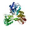
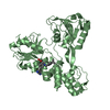
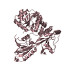
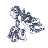
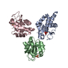
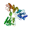
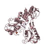
Entry Database : PDB / ID : 4wxpTitle X-ray crystal structure of NS3 Helicase from HCV with a bound fragment inhibitor at 2.08 A resolution NS3-4 protease Keywords / / Function / homology Function Domain/homology Component
/ / / / / / / / / / / / / / / / / / / / / / / / / / / / / / / / / / / / / / / / / / / / / / / / / / / / / / / / / / / / / / / / / / / / / / / / / / / / / / / / / / / / / / / / / / / / / / / / / / / / / / / / / / / / / / / / / / / / / / / / Biological species Method / / Resolution : 2.08 Å Authors Davies, D.R. Journal : TO BE PUBLISHED Title : crystal structure of NS3 Helicase from HCV with a bound fragment inhibitorAuthors: Buckman, B.O. / Kossen, K. / Misialek, S. / Stevens, S. / Rajagopalan, R. / Ruhmund, D. / Hooi, L. / Snarskaya, N. / Serebryany, V. / Wang, G. / Stoycheva, A. / Nicholas, J.B. / Davies, D.R. ... Authors : Buckman, B.O. / Kossen, K. / Misialek, S. / Stevens, S. / Rajagopalan, R. / Ruhmund, D. / Hooi, L. / Snarskaya, N. / Serebryany, V. / Wang, G. / Stoycheva, A. / Nicholas, J.B. / Davies, D.R. / Brunton, S. / Montalbetti, C. / Weddell, D. / Goodwin, C. / Schonfeld, D. / Mather, O. / Cheng, R. / Blatt, L. / Beigelman, L. History Deposition Nov 14, 2014 Deposition site / Processing site Revision 1.0 Dec 23, 2015 Provider / Type Revision 1.1 Sep 27, 2023 Group Data collection / Database references ... Data collection / Database references / Derived calculations / Refinement description Category chem_comp_atom / chem_comp_bond ... chem_comp_atom / chem_comp_bond / database_2 / diffrn_radiation_wavelength / pdbx_initial_refinement_model / pdbx_struct_conn_angle / pdbx_struct_oper_list / struct_conn Item _database_2.pdbx_DOI / _database_2.pdbx_database_accession ... _database_2.pdbx_DOI / _database_2.pdbx_database_accession / _pdbx_struct_conn_angle.ptnr1_auth_comp_id / _pdbx_struct_conn_angle.ptnr1_auth_seq_id / _pdbx_struct_conn_angle.ptnr1_label_asym_id / _pdbx_struct_conn_angle.ptnr1_label_atom_id / _pdbx_struct_conn_angle.ptnr1_label_comp_id / _pdbx_struct_conn_angle.ptnr1_label_seq_id / _pdbx_struct_conn_angle.ptnr3_auth_comp_id / _pdbx_struct_conn_angle.ptnr3_auth_seq_id / _pdbx_struct_conn_angle.ptnr3_label_asym_id / _pdbx_struct_conn_angle.ptnr3_label_atom_id / _pdbx_struct_conn_angle.ptnr3_label_comp_id / _pdbx_struct_conn_angle.ptnr3_label_seq_id / _pdbx_struct_conn_angle.value / _pdbx_struct_oper_list.symmetry_operation / _struct_conn.pdbx_dist_value / _struct_conn.ptnr1_auth_comp_id / _struct_conn.ptnr1_auth_seq_id / _struct_conn.ptnr1_label_asym_id / _struct_conn.ptnr1_label_atom_id / _struct_conn.ptnr1_label_comp_id / _struct_conn.ptnr1_label_seq_id / _struct_conn.ptnr2_auth_comp_id / _struct_conn.ptnr2_auth_seq_id / _struct_conn.ptnr2_label_asym_id / _struct_conn.ptnr2_label_atom_id / _struct_conn.ptnr2_label_comp_id / _struct_conn.ptnr2_symmetry
Show all Show less
 Movie
Movie Controller

 Yorodumi
 Open data
 Basic information
 Components
 Keywords
 Function and homology information
 Hepatitis C virus
 X-RAY DIFFRACTION /
  Authors
 Citation
 Structure visualization
 Downloads & links
 Other downloads
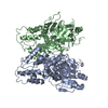


 PDBj
PDBj
 Assembly


 Mass: 22.990 Da / Num. of mol.: 1 / Source method: obtained synthetically / Formula: Na
 Mass: 35.453 Da / Num. of mol.: 1 / Source method: obtained synthetically / Formula: Cl
 Mass: 189.211 Da / Num. of mol.: 1 / Source method: obtained synthetically / Formula: C11H11NO2
 Mass: 18.015 Da / Num. of mol.: 117 / Source method: isolated from a natural source / Formula: H2O
 Sample preparation
 Processing