 Movie
Movie Controller

[English] 日本語
 Yorodumi
- PDB-6wp0: The Crystal Structure of Domain-Swapped Trimer Q108K:T51D variant... -
+ Open data

- Basic information
| Entry | Database: PDB / ID: 6wp0 | ||||||
|---|---|---|---|---|---|---|---|
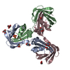
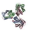
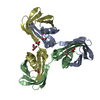
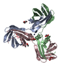
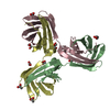
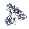
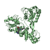
| Title | The Crystal Structure of Domain-Swapped Trimer Q108K:T51D variant of HCRBPII | ||||||
 Components Components | Retinol-binding protein 2 | ||||||
 Keywords Keywords | LIPID BINDING PROTEIN / iLBP | ||||||
| Function / homology |  Function and homology information Function and homology informationsynaptic ribbon / vitamin A metabolic process / all-trans-retinol binding / retinoid binding / retinal binding / molecular carrier activity / epidermis development / fatty acid transport / Retinoid metabolism and transport / retinoid metabolic process ...synaptic ribbon / vitamin A metabolic process / all-trans-retinol binding / retinoid binding / retinal binding / molecular carrier activity / epidermis development / fatty acid transport / Retinoid metabolism and transport / retinoid metabolic process / fatty acid binding / transmembrane transporter binding / nucleus / cytosol Similarity search - Function | ||||||
| Biological species |  Homo sapiens (human) Homo sapiens (human) | ||||||
| Method |  X-RAY DIFFRACTION / SYNCHROTRON / MOLECULAR REPLACEMENT / Resolution: 2.78 Å X-RAY DIFFRACTION / SYNCHROTRON / MOLECULAR REPLACEMENT / Resolution: 2.78 Å | ||||||
 Authors Authors | Ghanbarpour, A. / Geiger, J. | ||||||
| Funding support |  United States, 1items United States, 1items
| ||||||
 Citation Citation | Journal: Chembiochem / Year: 2020 Title: Human Cellular Retinol Binding Protein II Forms a Domain-Swapped Trimer Representing a Novel Fold and a New Template for Protein Engineering. Authors: Ghanbarpour, A. / Santos, E.M. / Pinger, C. / Assar, Z. / Hossaini Nasr, S. / Vasileiou, C. / Spence, D. / Borhan, B. / Geiger, J.H. #1: Journal: To Be PublishedTitle: The Crystal Structure of Apo Domain-Swapped Trimer Q108K:K40D:T53A:R58L:Q38F:Q4F variant of HCRBPII Authors: Ghanbarpour, A. / Geiger, J. | ||||||
| History |
|
- Structure visualization
| Structure viewer | Molecule: MolmilJmol/JSmol |
|---|
- Downloads & links
-Download
| PDBx/mmCIF format | 6wp0.cif.gz | 761.6 KB | Display | PDBx/mmCIF format |
|---|---|---|---|---|
| PDB format | pdb6wp0.ent.gz | 524.7 KB | Display | PDB format |
| PDBx/mmJSON format | 6wp0.json.gz | Tree view | PDBx/mmJSON format | |
| Others |  Other downloads Other downloads |
-Validation report
| Arichive directory | https://data.pdbj.org/pub/pdb/validation_reports/wp/6wp0ftp://data.pdbj.org/pub/pdb/validation_reports/wp/6wp0 | HTTPS FTP |
|---|
-Related structure data
| Related structure data |  6visC  6vitC 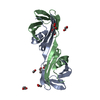 6wnfC  6wnjC 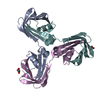 6wp1C  6wp2C  2rctS S: Starting model for refinement C: citing same article ( |
|---|---|
| Similar structure data |


-Links
 PDBj
PDBj


- Assembly
| Deposited unit | 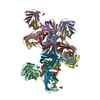
| |||||||||||||||||||||||||||||||||||||||||||||||||||||||||||||||||||||||||||||||||||||||||||||||||||||||||||
|---|---|---|---|---|---|---|---|---|---|---|---|---|---|---|---|---|---|---|---|---|---|---|---|---|---|---|---|---|---|---|---|---|---|---|---|---|---|---|---|---|---|---|---|---|---|---|---|---|---|---|---|---|---|---|---|---|---|---|---|---|---|---|---|---|---|---|---|---|---|---|---|---|---|---|---|---|---|---|---|---|---|---|---|---|---|---|---|---|---|---|---|---|---|---|---|---|---|---|---|---|---|---|---|---|---|---|---|---|
| 1 | 
| |||||||||||||||||||||||||||||||||||||||||||||||||||||||||||||||||||||||||||||||||||||||||||||||||||||||||||
| 2 | 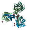
| |||||||||||||||||||||||||||||||||||||||||||||||||||||||||||||||||||||||||||||||||||||||||||||||||||||||||||
| 3 | 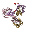
| |||||||||||||||||||||||||||||||||||||||||||||||||||||||||||||||||||||||||||||||||||||||||||||||||||||||||||
| 4 | 
| |||||||||||||||||||||||||||||||||||||||||||||||||||||||||||||||||||||||||||||||||||||||||||||||||||||||||||
| Unit cell |
| |||||||||||||||||||||||||||||||||||||||||||||||||||||||||||||||||||||||||||||||||||||||||||||||||||||||||||
| Noncrystallographic symmetry (NCS) | NCS domain:
NCS domain segments: Ens-ID: 1
|