 Movie
Movie Controller

[English] 日本語
 Yorodumi
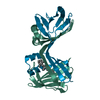
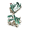
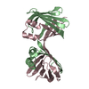
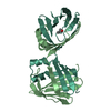
- PDB-6vit: The Crystal Structure of Apo Domain-Swapped dimer Q108K:T51D:I32C... -
+ Open data

- Basic information
| Entry | Database: PDB / ID: 6vit | ||||||
|---|---|---|---|---|---|---|---|
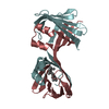
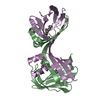
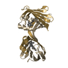
| Title | The Crystal Structure of Apo Domain-Swapped dimer Q108K:T51D:I32C Variant of HCRBPII with an Engineered Disulfide Bond | ||||||
 Components Components | Retinol-binding protein 2 | ||||||
 Keywords Keywords | LIPID BINDING PROTEIN / Domain Swapped Trimer / iLBP | ||||||
| Function / homology |  Function and homology information Function and homology informationsynaptic ribbon / vitamin A metabolic process / all-trans-retinol binding / retinoid binding / retinal binding / molecular carrier activity / epidermis development / fatty acid transport / Retinoid metabolism and transport / retinoid metabolic process ...synaptic ribbon / vitamin A metabolic process / all-trans-retinol binding / retinoid binding / retinal binding / molecular carrier activity / epidermis development / fatty acid transport / Retinoid metabolism and transport / retinoid metabolic process / fatty acid binding / transmembrane transporter binding / nucleus / cytosol Similarity search - Function | ||||||
| Biological species |  Homo sapiens (human) Homo sapiens (human) | ||||||
| Method |  X-RAY DIFFRACTION / SYNCHROTRON / MOLECULAR REPLACEMENT / Resolution: 3.2 Å X-RAY DIFFRACTION / SYNCHROTRON / MOLECULAR REPLACEMENT / Resolution: 3.2 Å | ||||||
 Authors Authors | Ghanbarpour, A. / Geiger, J. | ||||||
| Funding support |  United States, 1items United States, 1items
| ||||||
 Citation Citation | Journal: Chembiochem / Year: 2020 Title: Human Cellular Retinol Binding Protein II Forms a Domain-Swapped Trimer Representing a Novel Fold and a New Template for Protein Engineering. Authors: Ghanbarpour, A. / Santos, E.M. / Pinger, C. / Assar, Z. / Hossaini Nasr, S. / Vasileiou, C. / Spence, D. / Borhan, B. / Geiger, J.H. | ||||||
| History |
|
- Structure visualization
| Structure viewer | Molecule: MolmilJmol/JSmol |
|---|
- Downloads & links
-Download
| PDBx/mmCIF format | 6vit.cif.gz | 80.2 KB | Display | PDBx/mmCIF format |
|---|---|---|---|---|
| PDB format | pdb6vit.ent.gz | 49.3 KB | Display | PDB format |
| PDBx/mmJSON format | 6vit.json.gz | Tree view | PDBx/mmJSON format | |
| Others |  Other downloads Other downloads |
-Validation report
| Arichive directory | https://data.pdbj.org/pub/pdb/validation_reports/vi/6vitftp://data.pdbj.org/pub/pdb/validation_reports/vi/6vit | HTTPS FTP |
|---|
-Related structure data
| Related structure data | 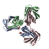 6visC 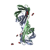 6wnfC  6wnjC  6wp0C 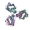 6wp1C  6wp2C  2rctS S: Starting model for refinement C: citing same article ( |
|---|---|
| Similar structure data |


-Links
 PDBj
PDBj


- Assembly
| Deposited unit | 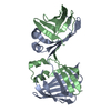
| |||||||||||||||||||||
|---|---|---|---|---|---|---|---|---|---|---|---|---|---|---|---|---|---|---|---|---|---|---|
| 1 |
| |||||||||||||||||||||
| Unit cell |
| |||||||||||||||||||||
| Noncrystallographic symmetry (NCS) | NCS domain:
NCS domain segments: Ens-ID: 1 / Beg auth comp-ID: THR / Beg label comp-ID: THR / End auth comp-ID: LYS / End label comp-ID: LYS / Auth seq-ID: 1 - 133 / Label seq-ID: 1 - 133
|
-Components
| #1: Protein | Mass: 15602.474 Da / Num. of mol.: 2 / Mutation: Q108K, T51D, I32C Source method: isolated from a genetically manipulated source Source: (gene. exp.) Homo sapiens (human) / Gene: RBP2, CRBP2Production host: Bacterial expression vector pET-11a (others) References: UniProt: P50120 #2: Water | ChemComp-HOH / |  Mass: 18.015 Da / Num. of mol.: 11 / Source method: isolated from a natural source / Formula: H2O Mass: 18.015 Da / Num. of mol.: 11 / Source method: isolated from a natural source / Formula: H2OHas protein modification | Y | |
|---|
-Experimental details
-Experiment
| Experiment | Method: X-RAY DIFFRACTION / Number of used crystals: 1 |
|---|
- Sample preparation
| Crystal | Density Matthews: 4.2 Å3/Da / Density % sol: 70.74 % |
|---|---|
| Crystal grow | Temperature: 298 K / Method: vapor diffusion, hanging drop / pH: 6.4 / Details: citric acid, PEG 3350 |
-Data collection
| Diffraction | Mean temperature: 100 K / Serial crystal experiment: N |
|---|---|
| Diffraction source | Source: SYNCHROTRON / Site: APS / Beamline: 21-ID-D / Wavelength: 0.97624 Å |
| Detector | Type: DECTRIS EIGER2 X 9M / Detector: PIXEL / Date: Nov 17, 2017 |
| Radiation | Protocol: SINGLE WAVELENGTH / Monochromatic (M) / Laue (L): M / Scattering type: x-ray |
| Radiation wavelength | Wavelength: 0.97624 Å / Relative weight: 1 |
| Reflection | Resolution: 3.2→50 Å / Num. obs: 8637 / % possible obs: 99.4 % / Redundancy: 5 % / Biso Wilson estimate: 78.05 Å2 / Rmerge(I) obs: 0.2 / Rrim(I) all: 0.21 / Net I/σ(I): 13.85 |
| Reflection shell | Resolution: 3.2→3.42 Å / Rmerge(I) obs: 1.1 / Mean I/σ(I) obs: 2.1 / Num. unique obs: 843 |
- Processing
| Software |
| |||||||||||||||||||||||||||||||||||||||||||||||||
|---|---|---|---|---|---|---|---|---|---|---|---|---|---|---|---|---|---|---|---|---|---|---|---|---|---|---|---|---|---|---|---|---|---|---|---|---|---|---|---|---|---|---|---|---|---|---|---|---|---|---|
| Refinement | Method to determine structure: MOLECULAR REPLACEMENT Starting model: 2rct Resolution: 3.2→38.48 Å / SU ML: 0.5048 / Cross valid method: FREE R-VALUE / σ(F): 1.36 / Phase error: 29.0483 / Stereochemistry target values: CDL v1.2
| |||||||||||||||||||||||||||||||||||||||||||||||||
| Solvent computation | Shrinkage radii: 0.9 Å / VDW probe radii: 1.11 Å / Solvent model: FLAT BULK SOLVENT MODEL | |||||||||||||||||||||||||||||||||||||||||||||||||
| Displacement parameters | Biso mean: 70.86 Å2 | |||||||||||||||||||||||||||||||||||||||||||||||||
| Refinement step | Cycle: LAST / Resolution: 3.2→38.48 Å
| |||||||||||||||||||||||||||||||||||||||||||||||||
| Refine LS restraints |
| |||||||||||||||||||||||||||||||||||||||||||||||||
| LS refinement shell |
|
