 Movie
Movie Controller

[English] 日本語
 Yorodumi
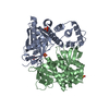
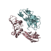
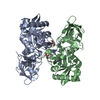
- PDB-4wx5: pore-forming thermostable direct hemolysin from Grimontia hollisae -
+ Open data

- Basic information
| Entry | Database: PDB / ID: 4wx5 | ||||||
|---|---|---|---|---|---|---|---|
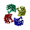
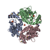
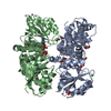
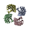
| Title | pore-forming thermostable direct hemolysin from Grimontia hollisae | ||||||
 Components Components | Hemolysin, heat labile | ||||||
 Keywords Keywords | TOXIN / thermostable direct hemolysin / TDH / oligomerization | ||||||
| Function / homology |  Function and homology information Function and homology informationsymbiont-mediated hemolysis of host erythrocyte / toxin activity / extracellular region Similarity search - Function | ||||||
| Biological species |  Grimontia hollisae (bacteria) Grimontia hollisae (bacteria) | ||||||
| Method |  X-RAY DIFFRACTION / SYNCHROTRON / MAD / Resolution: 2.302 Å X-RAY DIFFRACTION / SYNCHROTRON / MAD / Resolution: 2.302 Å | ||||||
 Authors Authors | Wang, Y.-K. / Wu, T.-K. / Li, T.-H.T. | ||||||
| Funding support |  Taiwan, 1items Taiwan, 1items
| ||||||
 Citation Citation | Journal: To be published Title: Multiple pleomorphic tetramers of pore-forming thermostable direct hemolysin from Grimontia hollisae in exerting membrane binding and hemolytic activity Authors: Wang, Y.-K. / Huang, S.-C. / Huang, W.-T. / Chang, C.-Y. / Kuo, T.-M. / Yip, B.-S. / Wu, T.-K. / Li, T.-H.T. | ||||||
| History |
|
- Structure visualization
| Structure viewer | Molecule: MolmilJmol/JSmol |
|---|
- Downloads & links
-Download
| PDBx/mmCIF format | 4wx5.cif.gz | 133.1 KB | Display | PDBx/mmCIF format |
|---|---|---|---|---|
| PDB format | pdb4wx5.ent.gz | 104.1 KB | Display | PDB format |
| PDBx/mmJSON format | 4wx5.json.gz | Tree view | PDBx/mmJSON format | |
| Others |  Other downloads Other downloads |
-Validation report
| Arichive directory | https://data.pdbj.org/pub/pdb/validation_reports/wx/4wx5ftp://data.pdbj.org/pub/pdb/validation_reports/wx/4wx5 | HTTPS FTP |
|---|
-Related structure data


-Links
 PDBj
PDBj- Assembly
| Deposited unit | 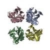
| ||||||||
|---|---|---|---|---|---|---|---|---|---|
| 1 |
| ||||||||
| Unit cell |
|
-Components
| #1: Protein | Mass: 18643.596 Da / Num. of mol.: 4 Source method: isolated from a genetically manipulated source Source: (gene. exp.) Grimontia hollisae (bacteria) / Plasmid: pCR2.1-TOPO / Production host: #2: Water | ChemComp-HOH / |  Mass: 18.015 Da / Num. of mol.: 184 / Source method: isolated from a natural source / Formula: H2O Mass: 18.015 Da / Num. of mol.: 184 / Source method: isolated from a natural source / Formula: H2OHas protein modification | Y | Sequence details | THE AUTHORS USED GRIMONTIA HOLLISAE STRAIN ATCC33564. THE AUTHORS ARE CONVINCED OF THIS SEQUENCE BY ...THE AUTHORS USED GRIMONTIA HOLLISAE STRAIN ATCC33564. THE AUTHORS ARE CONVINCED OF THIS SEQUENCE BY THE ELECTRON DENSITY MAP. | |
|---|
-Experimental details
-Experiment
| Experiment | Method: X-RAY DIFFRACTION / Number of used crystals: 1 |
|---|
- Sample preparation
| Crystal | Density Matthews: 2.64 Å3/Da / Density % sol: 53.38 % |
|---|---|
| Crystal grow | Temperature: 298 K / Method: vapor diffusion, hanging drop / pH: 7.5 Details: 28%(v/v) PEG 400, 0.2 M CaCl2, 0.1 M Na-HEPES buffer |
-Data collection
| Diffraction | Mean temperature: 100 K | ||||||||||||
|---|---|---|---|---|---|---|---|---|---|---|---|---|---|
| Diffraction source | Source: SYNCHROTRON / Site: NSRRC / Beamline: BL13C1 / Wavelength: 0.96379, 0.97904, 0.97920 | ||||||||||||
| Detector | Type: ADSC QUANTUM 315r / Detector: CCD / Date: Jan 15, 2010 | ||||||||||||
| Radiation | Protocol: MAD / Monochromatic (M) / Laue (L): M / Scattering type: x-ray | ||||||||||||
| Radiation wavelength |
| ||||||||||||
| Reflection | Resolution: 2.3→30 Å / Num. obs: 32500 / % possible obs: 99.6 % / Observed criterion σ(F): 0 / Observed criterion σ(I): -3 / Redundancy: 8.1 % / Rmerge(I) obs: 0.083 / Rsym value: 0.083 / Net I/σ(I): 21.8 | ||||||||||||
| Reflection shell | Resolution: 2.3→2.38 Å / Redundancy: 7.2 % / Rmerge(I) obs: 0.264 / Mean I/σ(I) obs: 6.7 / % possible all: 97.9 |
-Phasing
| Phasing | Method: MAD |
|---|
- Processing
| Software |
| |||||||||||||||||||||||||||||||||||||||||||||||||||||||||||||||||||||||||||||||||||||||||||||||||||||||||
|---|---|---|---|---|---|---|---|---|---|---|---|---|---|---|---|---|---|---|---|---|---|---|---|---|---|---|---|---|---|---|---|---|---|---|---|---|---|---|---|---|---|---|---|---|---|---|---|---|---|---|---|---|---|---|---|---|---|---|---|---|---|---|---|---|---|---|---|---|---|---|---|---|---|---|---|---|---|---|---|---|---|---|---|---|---|---|---|---|---|---|---|---|---|---|---|---|---|---|---|---|---|---|---|---|---|---|
| Refinement | Method to determine structure: MAD / Resolution: 2.302→26.768 Å / FOM work R set: 0.7285 / SU ML: 0.24 / Cross valid method: FREE R-VALUE / σ(F): 0 / Phase error: 31.48 / Stereochemistry target values: MLHL
| |||||||||||||||||||||||||||||||||||||||||||||||||||||||||||||||||||||||||||||||||||||||||||||||||||||||||
| Solvent computation | Shrinkage radii: 0.9 Å / VDW probe radii: 1.11 Å / Solvent model: FLAT BULK SOLVENT MODEL | |||||||||||||||||||||||||||||||||||||||||||||||||||||||||||||||||||||||||||||||||||||||||||||||||||||||||
| Displacement parameters | Biso max: 80.31 Å2 / Biso mean: 31.36 Å2 / Biso min: 10.95 Å2 | |||||||||||||||||||||||||||||||||||||||||||||||||||||||||||||||||||||||||||||||||||||||||||||||||||||||||
| Refinement step | Cycle: final / Resolution: 2.302→26.768 Å
| |||||||||||||||||||||||||||||||||||||||||||||||||||||||||||||||||||||||||||||||||||||||||||||||||||||||||
| Refine LS restraints |
| |||||||||||||||||||||||||||||||||||||||||||||||||||||||||||||||||||||||||||||||||||||||||||||||||||||||||
| LS refinement shell | Refine-ID: X-RAY DIFFRACTION / Total num. of bins used: 14
|
