 Movie
Movie Controller

[English] 日本語
 Yorodumi
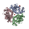
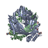
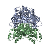
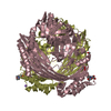
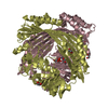
- PDB-1n3b: Crystal Structure of Dephosphocoenzyme A kinase from Escherichia coli -
+ Open data

- Basic information
| Entry | Database: PDB / ID: 1n3b | ||||||
|---|---|---|---|---|---|---|---|
| Title | Crystal Structure of Dephosphocoenzyme A kinase from Escherichia coli | ||||||
 Components Components | Dephospho-CoA kinase | ||||||
 Keywords Keywords | TRANSFERASE / trimer / P-loop / alpha/beta / Montreal-Kingston Bacterial Structural Genomics Initiative / BSGI / Structural Genomics | ||||||
| Function / homology |  Function and homology information Function and homology informationdephospho-CoA kinase / dephospho-CoA kinase activity / coenzyme A biosynthetic process / ATP binding / cytoplasm Similarity search - Function | ||||||
| Biological species |  | ||||||
| Method |  X-RAY DIFFRACTION / SYNCHROTRON / MAD / Resolution: 1.8 Å X-RAY DIFFRACTION / SYNCHROTRON / MAD / Resolution: 1.8 Å | ||||||
 Authors Authors | O'Toole, N. / Barbosa, J.A.R.G. / Li, Y. / Hung, L.-W. / Matte, A. / Cygler, M. / Montreal-Kingston Bacterial Structural Genomics Initiative (BSGI) | ||||||
 Citation Citation | Journal: Protein Sci. / Year: 2003 Title: Crystal Structure of a Trimeric Form of Dephosphocoenzyme A Kinase from Escherichia coli Authors: O'Toole, N. / Barbosa, J.A.R.G. / Li, Y. / Hung, L.-W. / Matte, A. / Cygler, M. #1: Journal: J.Struct.Biol. / Year: 2001Title: Crystal Structure of Dephospho-coenzyme A Kinase from Haemophilus influenzae Authors: Oblomova, G. / Teplyakov, A. / Bonander, N. / Eisenstein, E. / Howard, A. / Gilliland, G. #2: Journal: J.Bacteriol. / Year: 2001Title: Identification of yace (coaE) as the Structural Gene for Dephosphocoenzyme A Kinase in Escherichia coli K-12 Authors: Mishra, P.K. / Park, P.K. / Drueckhammer, D.G. #3: Journal: J.Mol.Biol. / Year: 1998Title: The structure of a Trimeric Archael Adenylate Kinase Authors: Vorhein, C. / Bonisch, H. / Schafer, G. / Schulz, G.E. #4: Journal: Biochem.J. / Year: 1983Title: A Bifunctional Enzyme Complex in Coenzyme A Biosynthesis: Purification of Pantetheine Phosphate Adenylyltransferase and Dephospho-CoA Kinase Authors: Worral, D.M. / Tubbs, P.K. #5: Journal: Biochem.J. / Year: 2002Title: Identification and Characterization of the Gene Encoding the Human Adenylyltransferase and Dephosphocoenzyme A Kinase Bifunctional Enzyme Authors: Aghajanian, S. / Worral, D.M. | ||||||
| History |
|
- Structure visualization
| Structure viewer | Molecule: MolmilJmol/JSmol |
|---|
- Downloads & links
-Download
| PDBx/mmCIF format | 1n3b.cif.gz | 136.1 KB | Display | PDBx/mmCIF format |
|---|---|---|---|---|
| PDB format | pdb1n3b.ent.gz | 106.8 KB | Display | PDB format |
| PDBx/mmJSON format | 1n3b.json.gz | Tree view | PDBx/mmJSON format | |
| Others |  Other downloads Other downloads |
-Validation report
| Arichive directory | https://data.pdbj.org/pub/pdb/validation_reports/n3/1n3bftp://data.pdbj.org/pub/pdb/validation_reports/n3/1n3b | HTTPS FTP |
|---|
-Related structure data
| Similar structure data | |
|---|---|
| Other databases |


-Links
 PDBj
PDBj- Assembly
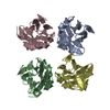
| Deposited unit | 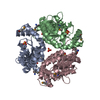
| ||||||||
|---|---|---|---|---|---|---|---|---|---|
| 1 |
| ||||||||
| Unit cell |
|
-Components
| #1: Protein | Mass: 24037.652 Da / Num. of mol.: 3 Source method: isolated from a genetically manipulated source Source: (gene. exp.) #2: Chemical | ChemComp-SO4 /   Mass: 96.063 Da / Num. of mol.: 10 / Source method: obtained synthetically / Formula: SO4 Mass: 96.063 Da / Num. of mol.: 10 / Source method: obtained synthetically / Formula: SO4#3: Water | ChemComp-HOH / |  Mass: 18.015 Da / Num. of mol.: 449 / Source method: isolated from a natural source / Formula: H2O Mass: 18.015 Da / Num. of mol.: 449 / Source method: isolated from a natural source / Formula: H2OHas protein modification | Y | |
|---|
-Experimental details
-Experiment
| Experiment | Method: X-RAY DIFFRACTION / Number of used crystals: 1 |
|---|
- Sample preparation
| Crystal | Density Matthews: 2.45 Å3/Da / Density % sol: 49.44 % | ||||||||||||||||||||||||||||||||||||||||||||||||||||||||||||||||||||||
|---|---|---|---|---|---|---|---|---|---|---|---|---|---|---|---|---|---|---|---|---|---|---|---|---|---|---|---|---|---|---|---|---|---|---|---|---|---|---|---|---|---|---|---|---|---|---|---|---|---|---|---|---|---|---|---|---|---|---|---|---|---|---|---|---|---|---|---|---|---|---|---|
| Crystal grow | Temperature: 293 K / Method: vapor diffusion, hanging drop / pH: 6.5 Details: PEG 8000, cacodylate buffer, ammonium sulfate, glycerol, pH 6.5, VAPOR DIFFUSION, HANGING DROP, temperature 293K | ||||||||||||||||||||||||||||||||||||||||||||||||||||||||||||||||||||||
| Crystal grow | *PLUS Temperature: 21 ℃ / pH: 7.5 | ||||||||||||||||||||||||||||||||||||||||||||||||||||||||||||||||||||||
| Components of the solutions | *PLUS
|
-Data collection
| Diffraction | Mean temperature: 100 K | |||||||||
|---|---|---|---|---|---|---|---|---|---|---|
| Diffraction source | Source: SYNCHROTRON / Site: NSLS  / Beamline: X8C / Wavelength: 0.97957, 0.97941 / Beamline: X8C / Wavelength: 0.97957, 0.97941 | |||||||||
| Detector | Type: ADSC QUANTUM 4 / Detector: CCD / Date: Jul 4, 2001 / Details: monochromating crystal | |||||||||
| Radiation | Monochromator: graphite / Protocol: MAD / Monochromatic (M) / Laue (L): M / Scattering type: x-ray | |||||||||
| Radiation wavelength |
| |||||||||
| Reflection | Resolution: 1.8→40 Å / Num. obs: 59512 / % possible obs: 86.3 % / Redundancy: 6.7 % / Rsym value: 0.065 / Net I/σ(I): 26.1 | |||||||||
| Reflection shell | Resolution: 1.81→1.88 Å / Num. unique all: 5096 / Rsym value: 0.439 / % possible all: 74.3 | |||||||||
| Reflection | *PLUS Lowest resolution: 40 Å / Num. obs: 56459 / % possible obs: 88.7 % / Num. measured all: 380533 / Rmerge(I) obs: 0.063 | |||||||||
| Reflection shell | *PLUS Highest resolution: 1.8 Å / Lowest resolution: 1.87 Å / % possible obs: 71.1 % / Rmerge(I) obs: 0.546 |
- Processing
| Software |
| ||||||||||||||||||||
|---|---|---|---|---|---|---|---|---|---|---|---|---|---|---|---|---|---|---|---|---|---|
| Refinement | Method to determine structure: MAD / Resolution: 1.8→40 Å / Isotropic thermal model: isotropic / Cross valid method: Free-R / σ(F): 0 / σ(I): 0 / Stereochemistry target values: Engh & Huber Details: Bulk solvent correction employed. The structure was refined using Friedel's pairs.
| ||||||||||||||||||||
| Displacement parameters | Biso mean: 28.46 Å2 | ||||||||||||||||||||
| Refinement step | Cycle: LAST / Resolution: 1.8→40 Å
| ||||||||||||||||||||
| Refine LS restraints |
| ||||||||||||||||||||
| Refinement | *PLUS Highest resolution: 1.8 Å / Lowest resolution: 40 Å | ||||||||||||||||||||
| Solvent computation | *PLUS | ||||||||||||||||||||
| Displacement parameters | *PLUS | ||||||||||||||||||||
| Refine LS restraints | *PLUS Type: c_bond_d / Dev ideal: 0.008 |
