- PDB-4wws: Structure of Chlorite dismutase-like Protein from Listeria monocy... -
+
Open data
ID or keywords:
Loading...
-
Basic information
Entry
Database: PDB / ID: 4wws
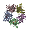
Title
Structure of Chlorite dismutase-like Protein from Listeria monocytogenes
Components
Putative heme-dependent peroxidase lmo2113
Keywords
OXIDOREDUCTASE / Ferredoxin-like fold / Chlorite dismutase-like protein
Function / homology
Function and homology information
oxidoreductase activity, acting on the CH-CH group of donors, oxygen as acceptor / hydrogen peroxide-dependent heme synthase / heme B biosynthetic process / peroxidase activity / heme binding / metal ion binding Similarity search - Function
Mass: 18.015 Da / Num. of mol.: 1020 / Source method: isolated from a natural source / Formula: H2O
-
Experimental details
-
Experiment
Experiment
Method: X-RAY DIFFRACTION / Number of used crystals: 1
-
Sample preparation
Crystal
Density Matthews: 2.59 Å3/Da / Density % sol: 52.56 % / Description: twin l,-k,h
Crystal grow
Temperature: 295 K / Method: vapor diffusion, hanging drop / pH: 9 Details: 1.1 M Na K tartrate, 8% glycerol, 0.1 M TrisHCl, 1/10 Silver Bullets 52: Protamine Sulfate
-
Data collection
Diffraction
ID
Mean temperature (K)
Crystal-ID
1
100
1
2
1
Diffraction source
Source
Site
Beamline
ID
Wavelength (Å)
SYNCHROTRON
ESRF
ID14-1
1
0.91841
SYNCHROTRON
ESRF
ID23-1
2
Detector
Type: MARMOSAIC 225 mm CCD / Detector: CCD / Date: Mar 3, 2012
Radiation
Protocol: SINGLE WAVELENGTH / Monochromatic (M) / Laue (L): M / Scattering type: x-ray
Radiation wavelength
Wavelength: 0.91841 Å / Relative weight: 1
Reflection twin
Operator: l,-k,h / Fraction: 0.26
Reflection
Resolution: 2→48.76 Å / Num. obs: 99373 / % possible obs: 99.8 % / Redundancy: 4.2 % / Rmerge(I) obs: 0.196 / Net I/σ(I): 6.77
Reflection shell
Resolution: 2→2.071 Å / Redundancy: 4.3 % / Rmerge(I) obs: 0.9392 / Mean I/σ(I) obs: 1.59 / % possible all: 99.99
-
Phasing
Phasing
Method: molecular replacement
-
Processing
Software
Name
Version
Classification
PHENIX
refinement
PDB_EXTRACT
3.15
dataextraction
XSCALE
datascaling
BALBES
phasing
XDS
datascaling
Refinement
Method to determine structure: MOLECULAR REPLACEMENT Starting model: 3NN1
In the structure databanks used in Yorodumi, some data are registered as the other names, "COVID-19 virus" and "2019-nCoV". Here are the details of the virus and the list of structure data.
Jan 31, 2019. EMDB accession codes are about to change! (news from PDBe EMDB page)
EMDB accession codes are about to change! (news from PDBe EMDB page)
The allocation of 4 digits for EMDB accession codes will soon come to an end. Whilst these codes will remain in use, new EMDB accession codes will include an additional digit and will expand incrementally as the available range of codes is exhausted. The current 4-digit format prefixed with “EMD-” (i.e. EMD-XXXX) will advance to a 5-digit format (i.e. EMD-XXXXX), and so on. It is currently estimated that the 4-digit codes will be depleted around Spring 2019, at which point the 5-digit format will come into force.
The EM Navigator/Yorodumi systems omit the EMD- prefix.
Related info.:Q: What is EMD? / ID/Accession-code notation in Yorodumi/EM Navigator
Yorodumi is a browser for structure data from EMDB, PDB, SASBDB, etc.
This page is also the successor to EM Navigator detail page, and also detail information page/front-end page for Omokage search.
The word "yorodu" (or yorozu) is an old Japanese word meaning "ten thousand". "mi" (miru) is to see.
Related info.:EMDB / PDB / SASBDB / Comparison of 3 databanks / Yorodumi Search / Aug 31, 2016. New EM Navigator & Yorodumi / Yorodumi Papers / Jmol/JSmol / Function and homology information / Changes in new EM Navigator and Yorodumi
 Movie
Movie Controller

 Yorodumi
 Open data
 Basic information
 Components
 Keywords
 Function and homology information
 Listeria monocytogenes serovar 1/2a (bacteria)
 X-RAY DIFFRACTION /
  Authors
 Citation
 Structure visualization
 Downloads & links
 Other downloads
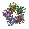


 PDBj
PDBj Assembly

 Mass: 39.098 Da / Num. of mol.: 5 / Source method: obtained synthetically / Formula: K
 Mass: 18.015 Da / Num. of mol.: 1020 / Source method: isolated from a natural source / Formula: H2O
 Sample preparation
 Processing