 Movie
Movie Controller

[English] 日本語
 Yorodumi
- PDB-5t2k: Geobacillus stearothermophilus HemQ with Manganese-Coproporphyrin III -
+ Open data

- Basic information
| Entry | Database: PDB / ID: 5t2k | ||||||
|---|---|---|---|---|---|---|---|
| Title | Geobacillus stearothermophilus HemQ with Manganese-Coproporphyrin III | ||||||
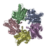
 Components Components | Putative heme-dependent peroxidase GT50_08830 | ||||||
 Keywords Keywords | OXIDOREDUCTASE / HemQ / Coproporphyrin III / Decarboxylation | ||||||
| Function / homology |  Function and homology information Function and homology informationhydrogen peroxide-dependent heme synthase / heme biosynthetic process / peroxidase activity / heme binding / metal ion binding Similarity search - Function | ||||||
| Biological species |   Geobacillus stearothermophilus 10 (bacteria) Geobacillus stearothermophilus 10 (bacteria) | ||||||
| Method |  X-RAY DIFFRACTION / SYNCHROTRON / MOLECULAR REPLACEMENT / Resolution: 1.8 Å X-RAY DIFFRACTION / SYNCHROTRON / MOLECULAR REPLACEMENT / Resolution: 1.8 Å | ||||||
 Authors Authors | Gauss, G.H. / Celis, A.I. / Dubois, J.L. / Peters, J.W. | ||||||
 Citation Citation | Journal: J. Am. Chem. Soc. / Year: 2017 Title: Structure-Based Mechanism for Oxidative Decarboxylation Reactions Mediated by Amino Acids and Heme Propionates in Coproheme Decarboxylase (HemQ). Authors: Celis, A.I. / Gauss, G.H. / Streit, B.R. / Shisler, K. / Moraski, G.C. / Rodgers, K.R. / Lukat-Rodgers, G.S. / Peters, J.W. / DuBois, J.L. | ||||||
| History |
|
- Structure visualization
| Structure viewer | Molecule: MolmilJmol/JSmol |
|---|
- Downloads & links
-Download
| PDBx/mmCIF format | 5t2k.cif.gz | 505.7 KB | Display | PDBx/mmCIF format |
|---|---|---|---|---|
| PDB format | pdb5t2k.ent.gz | 423.3 KB | Display | PDB format |
| PDBx/mmJSON format | 5t2k.json.gz | Tree view | PDBx/mmJSON format | |
| Others |  Other downloads Other downloads |
-Validation report
| Arichive directory | https://data.pdbj.org/pub/pdb/validation_reports/t2/5t2kftp://data.pdbj.org/pub/pdb/validation_reports/t2/5t2k | HTTPS FTP |
|---|
-Related structure data
| Related structure data |  1t0tS S: Starting model for refinement |
|---|---|
| Similar structure data |


-Links
 PDBj
PDBj- Assembly
| Deposited unit | 
| ||||||||
|---|---|---|---|---|---|---|---|---|---|
| 1 |
| ||||||||
| Unit cell |
|
-Components
| #1: Protein | Mass: 28817.609 Da / Num. of mol.: 5 Source method: isolated from a genetically manipulated source Source: (gene. exp.) Geobacillus stearothermophilus 10 (bacteria)Gene: GT50_08830 / Production host: References: UniProt: A0A0K2H9D8, Oxidoreductases; Acting on a peroxide as acceptor; Peroxidases #2: Chemical | ChemComp-76R / [   Mass: 707.631 Da / Num. of mol.: 5 / Source method: obtained synthetically / Formula: C36H36MnN4O8 Mass: 707.631 Da / Num. of mol.: 5 / Source method: obtained synthetically / Formula: C36H36MnN4O8#3: Water | ChemComp-HOH / |  Mass: 18.015 Da / Num. of mol.: 1373 / Source method: isolated from a natural source / Formula: H2O Mass: 18.015 Da / Num. of mol.: 1373 / Source method: isolated from a natural source / Formula: H2O |
|---|
-Experimental details
-Experiment
| Experiment | Method: X-RAY DIFFRACTION / Number of used crystals: 1 |
|---|
- Sample preparation
| Crystal | Density Matthews: 2.72 Å3/Da / Density % sol: 54.71 % |
|---|---|
| Crystal grow | Temperature: 293 K / Method: vapor diffusion, hanging drop / pH: 7.5 Details: 0.1 M HEPES pH 7.5, 0.05 M cadmium sulfate, 0.8-1.2 M sodium acetate |
-Data collection
| Diffraction | Mean temperature: 100 K |
|---|---|
| Diffraction source | Source: SYNCHROTRON / Site: SSRL  / Beamline: BL14-1 / Wavelength: 1 Å / Beamline: BL14-1 / Wavelength: 1 Å |
| Detector | Type: MARMOSAIC 325 mm CCD / Detector: CCD / Date: Jun 26, 2016 |
| Radiation | Protocol: SINGLE WAVELENGTH / Monochromatic (M) / Laue (L): M / Scattering type: x-ray |
| Radiation wavelength | Wavelength: 1 Å / Relative weight: 1 |
| Reflection | Resolution: 1.8→38.4 Å / Num. obs: 142652 / % possible obs: 99.3 % / Redundancy: 3.5 % / CC1/2: 0.998 / Rmerge(I) obs: 0.057 / Net I/σ(I): 13 |
| Reflection shell | Resolution: 1.8→1.83 Å / Redundancy: 3.3 % / Rmerge(I) obs: 0.514 / Mean I/σ(I) obs: 2.1 / CC1/2: 0.824 / % possible all: 86.7 |
- Processing
| Software |
| |||||||||||||||||||||||||||||||||||||||||||||||||||||||||||||||||||||||||||||||||||||||||||||||||||||||||||||||||||||||||||||||||||||||||||||||||||||||||||||||||||||||||||||||||||||||||||||||||||||||||||||||||||||||||
|---|---|---|---|---|---|---|---|---|---|---|---|---|---|---|---|---|---|---|---|---|---|---|---|---|---|---|---|---|---|---|---|---|---|---|---|---|---|---|---|---|---|---|---|---|---|---|---|---|---|---|---|---|---|---|---|---|---|---|---|---|---|---|---|---|---|---|---|---|---|---|---|---|---|---|---|---|---|---|---|---|---|---|---|---|---|---|---|---|---|---|---|---|---|---|---|---|---|---|---|---|---|---|---|---|---|---|---|---|---|---|---|---|---|---|---|---|---|---|---|---|---|---|---|---|---|---|---|---|---|---|---|---|---|---|---|---|---|---|---|---|---|---|---|---|---|---|---|---|---|---|---|---|---|---|---|---|---|---|---|---|---|---|---|---|---|---|---|---|---|---|---|---|---|---|---|---|---|---|---|---|---|---|---|---|---|---|---|---|---|---|---|---|---|---|---|---|---|---|---|---|---|---|---|---|---|---|---|---|---|---|---|---|---|---|---|---|---|---|
| Refinement | Method to determine structure: MOLECULAR REPLACEMENT Starting model: 1T0T Resolution: 1.8→38.4 Å / SU ML: 0.16 / Cross valid method: THROUGHOUT / σ(F): 1.35 / Phase error: 18.28
| |||||||||||||||||||||||||||||||||||||||||||||||||||||||||||||||||||||||||||||||||||||||||||||||||||||||||||||||||||||||||||||||||||||||||||||||||||||||||||||||||||||||||||||||||||||||||||||||||||||||||||||||||||||||||
| Solvent computation | Shrinkage radii: 0.9 Å / VDW probe radii: 1.11 Å | |||||||||||||||||||||||||||||||||||||||||||||||||||||||||||||||||||||||||||||||||||||||||||||||||||||||||||||||||||||||||||||||||||||||||||||||||||||||||||||||||||||||||||||||||||||||||||||||||||||||||||||||||||||||||
| Refinement step | Cycle: LAST / Resolution: 1.8→38.4 Å
| |||||||||||||||||||||||||||||||||||||||||||||||||||||||||||||||||||||||||||||||||||||||||||||||||||||||||||||||||||||||||||||||||||||||||||||||||||||||||||||||||||||||||||||||||||||||||||||||||||||||||||||||||||||||||
| Refine LS restraints |
| |||||||||||||||||||||||||||||||||||||||||||||||||||||||||||||||||||||||||||||||||||||||||||||||||||||||||||||||||||||||||||||||||||||||||||||||||||||||||||||||||||||||||||||||||||||||||||||||||||||||||||||||||||||||||
| LS refinement shell |
|
