 Movie
Movie Controller

[English] 日本語
 Yorodumi
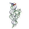
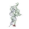
- PDB-4wul: Crystal structure of E. faecalis DNA binding domain LiaRD191N com... -
+ Open data

- Basic information
| Entry | Database: PDB / ID: 4wul | ||||||
|---|---|---|---|---|---|---|---|
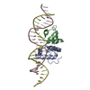
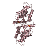
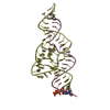
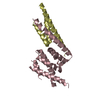
| Title | Crystal structure of E. faecalis DNA binding domain LiaRD191N complexed with 26bp DNA | ||||||
 Components Components |
| ||||||
 Keywords Keywords | DNA BINDING PROTEIN/DNA / helix-turn-helix / response regulator / enterococci / DNA binding domain / DNA BINDING PROTEIN-DNA complex | ||||||
| Function / homology |  Function and homology information Function and homology informationphosphorelay signal transduction system / regulation of DNA-templated transcription / DNA binding Similarity search - Function | ||||||
| Biological species |  Enterococcus faecalis S613 (bacteria) Enterococcus faecalis S613 (bacteria) | ||||||
| Method |  X-RAY DIFFRACTION / SYNCHROTRON / MOLECULAR REPLACEMENT / molecular replacement / Resolution: 2.4 Å X-RAY DIFFRACTION / SYNCHROTRON / MOLECULAR REPLACEMENT / molecular replacement / Resolution: 2.4 Å | ||||||
 Authors Authors | Davlieva, M. / Shamoo, Y. | ||||||
| Funding support |  United States, 1items United States, 1items
| ||||||
 Citation Citation | Journal: Nucleic Acids Res. / Year: 2015 Title: A variable DNA recognition site organization establishes the LiaR-mediated cell envelope stress response of enterococci to daptomycin. Authors: Davlieva, M. / Shi, Y. / Leonard, P.G. / Johnson, T.A. / Zianni, M.R. / Arias, C.A. / Ladbury, J.E. / Shamoo, Y. | ||||||
| History |
|
- Structure visualization
| Structure viewer | Molecule: MolmilJmol/JSmol |
|---|
- Downloads & links
-Download
| PDBx/mmCIF format | 4wul.cif.gz | 122.3 KB | Display | PDBx/mmCIF format |
|---|---|---|---|---|
| PDB format | pdb4wul.ent.gz | 91.2 KB | Display | PDB format |
| PDBx/mmJSON format | 4wul.json.gz | Tree view | PDBx/mmJSON format | |
| Others |  Other downloads Other downloads |
-Validation report
| Arichive directory | https://data.pdbj.org/pub/pdb/validation_reports/wu/4wulftp://data.pdbj.org/pub/pdb/validation_reports/wu/4wul | HTTPS FTP |
|---|
-Related structure data
| Related structure data | 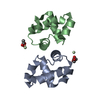 4wszC 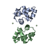 4wt0C 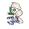 4wu4C 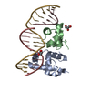 4wuhC  4wto C: citing same article ( S: Starting model for refinement |
|---|---|
| Similar structure data |


-Links
 PDBj
PDBj


- Assembly
| Deposited unit | 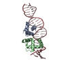
| ||||||||
|---|---|---|---|---|---|---|---|---|---|
| 1 |
| ||||||||
| Unit cell |
|
-Components
| #1: Protein | Mass: 7710.860 Da / Num. of mol.: 2 / Fragment: DNA binding domain Source method: isolated from a genetically manipulated source Plasmid: pETDuet / Production host: #2: DNA chain | | Mass: 8003.219 Da / Num. of mol.: 1 / Source method: obtained synthetically / Source: (synth.) Enterococcus faecalis S613 (bacteria)#3: DNA chain | | Mass: 7967.162 Da / Num. of mol.: 1 / Source method: obtained synthetically / Source: (synth.) Enterococcus faecalis S613 (bacteria)#4: Water | ChemComp-HOH / |  Mass: 18.015 Da / Num. of mol.: 98 / Source method: isolated from a natural source / Formula: H2O Mass: 18.015 Da / Num. of mol.: 98 / Source method: isolated from a natural source / Formula: H2O |
|---|
-Experimental details
-Experiment
| Experiment | Method: X-RAY DIFFRACTION / Number of used crystals: 1 |
|---|
- Sample preparation
| Crystal | Density Matthews: 2.93 Å3/Da / Density % sol: 57.99 % |
|---|---|
| Crystal grow | Temperature: 283 K / Method: vapor diffusion, hanging drop / pH: 8.6 Details: 0.08 M KCl, 0.02 M BaCl2, 0.04 M Sodium cacodylate (pH 6.0), 43% MPD, 0.012 M Spermine tetrachloride, 0.01 M Proline, 0.01 M Strontium chloride |
-Data collection
| Diffraction | Mean temperature: 100 K |
|---|---|
| Diffraction source | Source: SYNCHROTRON / Site: APS / Beamline: 21-ID-D / Wavelength: 0.97872 Å |
| Detector | Type: MARMOSAIC 300 mm CCD / Detector: CCD / Date: Dec 1, 2013 / Details: Bimorph K-B pair |
| Radiation | Monochromator: Kohzu / Protocol: SINGLE WAVELENGTH / Monochromatic (M) / Laue (L): M / Scattering type: x-ray |
| Radiation wavelength | Wavelength: 0.97872 Å / Relative weight: 1 |
| Reflection | Resolution: 2.4→30 Å / % possible obs: 100 % / Observed criterion σ(F): 0 / Observed criterion σ(I): -3 / Redundancy: 22.5 % / Rmerge(I) obs: 0.101 / Rsym value: 0.101 / Net I/av σ(I): 33.67 / Net I/σ(I): 33 |
| Reflection shell | Resolution: 2.4→2.4 Å / Redundancy: 22.3 % / Rmerge(I) obs: 0.733 / Mean I/σ(I) obs: 9.333 / Rsym value: 0.733 / % possible all: 100 |
| Cell measurement | Reflection used: 280748 |
-Phasing
| Phasing | Method: molecular replacement |
|---|
- Processing
| Software |
| |||||||||||||||||||||||||||||||||||||||||||||||||||||||||||||||||||||||||||||
|---|---|---|---|---|---|---|---|---|---|---|---|---|---|---|---|---|---|---|---|---|---|---|---|---|---|---|---|---|---|---|---|---|---|---|---|---|---|---|---|---|---|---|---|---|---|---|---|---|---|---|---|---|---|---|---|---|---|---|---|---|---|---|---|---|---|---|---|---|---|---|---|---|---|---|---|---|---|---|
| Refinement | Method to determine structure: MOLECULAR REPLACEMENT Starting model: 4WTO 4wto Resolution: 2.4→29.445 Å / SU ML: 0.34 / Cross valid method: FREE R-VALUE / σ(F): 1.34 / Phase error: 28.13 / Stereochemistry target values: ML
| |||||||||||||||||||||||||||||||||||||||||||||||||||||||||||||||||||||||||||||
| Solvent computation | Shrinkage radii: 0.9 Å / VDW probe radii: 1.11 Å / Solvent model: FLAT BULK SOLVENT MODEL | |||||||||||||||||||||||||||||||||||||||||||||||||||||||||||||||||||||||||||||
| Displacement parameters | Biso max: 162.9 Å2 / Biso mean: 46.9647 Å2 / Biso min: 10.7 Å2 | |||||||||||||||||||||||||||||||||||||||||||||||||||||||||||||||||||||||||||||
| Refinement step | Cycle: final / Resolution: 2.4→29.445 Å
| |||||||||||||||||||||||||||||||||||||||||||||||||||||||||||||||||||||||||||||
| Refine LS restraints |
| |||||||||||||||||||||||||||||||||||||||||||||||||||||||||||||||||||||||||||||
| LS refinement shell | Refine-ID: X-RAY DIFFRACTION / Total num. of bins used: 10
| |||||||||||||||||||||||||||||||||||||||||||||||||||||||||||||||||||||||||||||
| Refinement TLS params. | Method: refined / Origin x: 3.3254 Å / Origin y: 42.4077 Å / Origin z: -9.1498 Å
| |||||||||||||||||||||||||||||||||||||||||||||||||||||||||||||||||||||||||||||
| Refinement TLS group |
|
