Spanish Ministry of Economy and Competitiveness and the European Regional Development Fund
BFU2009-08389
スペイン
Spanish Ministry of Economy and Competitiveness and the European Regional Development Fund
BFU2012-32847
スペイン
引用
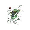
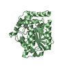
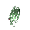
ジャーナル: Acta Crystallogr D Biol Crystallogr / 年: 2015 タイトル: Combination of X-ray crystallography, SAXS and DEER to obtain the structure of the FnIII-3,4 domains of integrin α6β4. 著者: Noelia Alonso-García / Inés García-Rubio / José A Manso / Rubén M Buey / Hector Urien / Arnoud Sonnenberg / Gunnar Jeschke / José M de Pereda / 要旨: Integrin α6β4 is a major component of hemidesmosomes that mediate the stable anchorage of epithelial cells to the underlying basement membrane. Integrin α6β4 has also been implicated in cell ...Integrin α6β4 is a major component of hemidesmosomes that mediate the stable anchorage of epithelial cells to the underlying basement membrane. Integrin α6β4 has also been implicated in cell proliferation and migration and in carcinoma progression. The third and fourth fibronectin type III domains (FnIII-3,4) of integrin β4 mediate binding to the hemidesmosomal proteins BPAG1e and BPAG2, and participate in signalling. Here, it is demonstrated that X-ray crystallography, small-angle X-ray scattering and double electron-electron resonance (DEER) complement each other to solve the structure of the FnIII-3,4 region. The crystal structures of the individual FnIII-3 and FnIII-4 domains were solved and the relative arrangement of the FnIII domains was elucidated by combining DEER with site-directed spin labelling. Multiple structures of the interdomain linker were modelled by Monte Carlo methods complying with DEER constraints, and the final structures were selected against experimental scattering data. FnIII-3,4 has a compact and cambered flat structure with an evolutionary conserved surface that is likely to correspond to a protein-interaction site. Finally, this hybrid method is of general application for the study of other macromolecules and complexes.
履歴
登録
2014年10月30日
登録サイト: RCSB / 処理サイト: PDBE
改定 1.0
2015年2月11日
Provider: repository / タイプ: Initial release
改定 1.1
2015年4月8日
Group: Database references
改定 1.2
2015年4月22日
Group: Database references
改定 2.0
2024年1月10日
Group: Atomic model / Data collection ...Atomic model / Data collection / Database references / Refinement description カテゴリ: atom_site / chem_comp_atom ...atom_site / chem_comp_atom / chem_comp_bond / database_2 / pdbx_initial_refinement_model Item: _atom_site.occupancy / _database_2.pdbx_DOI / _database_2.pdbx_database_accession
 ムービー
ムービー コントローラー


 データを開く
 基本情報
基本情報 要素
要素 キーワード
キーワード 機能・相同性情報
 Homo sapiens (ヒト)
 X線回折 /
  データ登録者
 スペイン, 2件
  引用
引用

 構造の表示
構造の表示 ダウンロードとリンク
 その他のダウンロード


 PDBj
PDBj


 集合体
集合体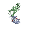


 分子量: 96.063 Da / 分子数: 2 / 由来タイプ: 合成 / 式: SO4
 分子量: 238.278 Da / 分子数: 1 / 由来タイプ: 合成 / 式: C10H22O6 / コメント: 沈殿剤*YM
 分子量: 62.068 Da / 分子数: 1 / 由来タイプ: 合成 / 式: C2H6O2
 分子量: 18.015 Da / 分子数: 219 / 由来タイプ: 天然 / 式: H2O
 試料調製
試料調製 解析
解析