 Movie
Movie Controller


+ Open data

- Basic information
| Entry | Database: PDB / ID: 4wrm | ||||||
|---|---|---|---|---|---|---|---|
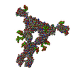
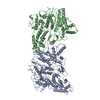
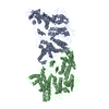
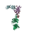
| Title | Structure of the human CSF-1:CSF-1R complex | ||||||
 Components Components |
| ||||||
 Keywords Keywords | CYTOKINE/CYTOKINE RECEPTOR / cytokine-cytokine receptor complex | ||||||
| Function / homology |  Function and homology information Function and homology informationregulation of macrophage derived foam cell differentiation / mammary gland fat development / positive regulation of macrophage colony-stimulating factor signaling pathway / macrophage homeostasis / macrophage colony-stimulating factor receptor binding / monocyte homeostasis / osteoclast proliferation / positive regulation of macrophage migration / positive regulation of odontogenesis of dentin-containing tooth / macrophage colony-stimulating factor receptor activity ...regulation of macrophage derived foam cell differentiation / mammary gland fat development / positive regulation of macrophage colony-stimulating factor signaling pathway / macrophage homeostasis / macrophage colony-stimulating factor receptor binding / monocyte homeostasis / osteoclast proliferation / positive regulation of macrophage migration / positive regulation of odontogenesis of dentin-containing tooth / macrophage colony-stimulating factor receptor activity / CSF1-CSF1R complex / forebrain neuron differentiation / monocyte activation / macrophage colony-stimulating factor signaling pathway / developmental process involved in reproduction / regulation of macrophage migration / positive regulation of microglial cell migration / mammary duct terminal end bud growth / cellular response to macrophage colony-stimulating factor stimulus / positive regulation of protein tyrosine kinase activity / microglial cell proliferation / positive regulation of mononuclear cell proliferation / cell-cell junction maintenance / olfactory bulb development / positive regulation of macrophage differentiation / mammary gland duct morphogenesis / myeloid leukocyte migration / host-mediated activation of viral process / ruffle organization / positive regulation of monocyte differentiation / neutrophil homeostasis / positive regulation of multicellular organism growth / positive regulation of osteoclast differentiation / branching involved in mammary gland duct morphogenesis / positive regulation of macrophage derived foam cell differentiation / positive regulation of macrophage proliferation / positive regulation of Ras protein signal transduction / regulation of bone resorption / Other interleukin signaling / positive regulation of cell motility / positive regulation of tyrosine phosphorylation of STAT protein / positive regulation of cell-matrix adhesion / growth factor binding / positive regulation of macrophage chemotaxis / cellular response to cytokine stimulus / cytokine binding / Interleukin-10 signaling / positive regulation of protein metabolic process / Transcriptional Regulation by VENTX / macrophage differentiation / regulation of MAPK cascade / hemopoiesis / regulation of ossification / monocyte differentiation / positive regulation of chemokine production / peptidyl-tyrosine phosphorylation / Transcriptional and post-translational regulation of MITF-M expression and activity / osteoclast differentiation / homeostasis of number of cells within a tissue / cell surface receptor protein tyrosine kinase signaling pathway / cytokine activity / axon guidance / response to ischemia / regulation of actin cytoskeleton organization / growth factor activity / Post-translational protein phosphorylation / receptor protein-tyrosine kinase / positive regulation of protein phosphorylation / Regulation of Insulin-like Growth Factor (IGF) transport and uptake by Insulin-like Growth Factor Binding Proteins (IGFBPs) / cytokine-mediated signaling pathway / Signaling by CSF1 (M-CSF) in myeloid cells / protein autophosphorylation / regulation of cell shape / cell migration / protein tyrosine kinase activity / protein phosphatase binding / Ras protein signal transduction / positive regulation of ERK1 and ERK2 cascade / positive regulation of phosphatidylinositol 3-kinase/protein kinase B signal transduction / signaling receptor complex / cell population proliferation / nuclear body / positive regulation of cell migration / endoplasmic reticulum lumen / inflammatory response / negative regulation of cell population proliferation / innate immune response / positive regulation of cell population proliferation / positive regulation of gene expression / negative regulation of apoptotic process / perinuclear region of cytoplasm / cell surface / signal transduction / protein homodimerization activity / : / extracellular region / nucleoplasm / ATP binding / membrane / identical protein binding Similarity search - Function | ||||||
| Biological species |  Homo sapiens (human) Homo sapiens (human) | ||||||
| Method |  X-RAY DIFFRACTION / SYNCHROTRON / MOLECULAR REPLACEMENT / Resolution: 6.853 Å X-RAY DIFFRACTION / SYNCHROTRON / MOLECULAR REPLACEMENT / Resolution: 6.853 Å | ||||||
 Authors Authors | Felix, J. / De Munck, S. / Elegheert, J. / Savvides, S.N. | ||||||
 Citation Citation | Journal: Structure / Year: 2015 Title: Structure and Assembly Mechanism of the Signaling Complex Mediated by Human CSF-1. Authors: Jan Felix / Steven De Munck / Kenneth Verstraete / Leander Meuris / Nico Callewaert / Jonathan Elegheert / Savvas N Savvides /  Abstract: Human colony-stimulating factor 1 receptor (hCSF-1R) is unique among the hematopoietic receptors because it is activated by two distinct cytokines, CSF-1 and interleukin-34 (IL-34). Despite ever- ...Human colony-stimulating factor 1 receptor (hCSF-1R) is unique among the hematopoietic receptors because it is activated by two distinct cytokines, CSF-1 and interleukin-34 (IL-34). Despite ever-growing insights into the central role of hCSF-1R signaling in innate and adaptive immunity, inflammatory diseases, and cancer, the structural basis of the functional dichotomy of hCSF-1R has remained elusive. Here, we report crystal structures of ternary complexes between hCSF-1 and hCSF-1R, including their complete extracellular assembly, and propose a mechanism for the cooperative human CSF-1:CSF-1R complex that relies on the adoption by dimeric hCSF-1 of an active conformational state and homotypic receptor interactions. Furthermore, we trace the cytokine-binding duality of hCSF-1R to a limited set of conserved interactions mediated by functionally equivalent residues on CSF-1 and IL-34 that play into the geometric requirements of hCSF-1R activation, and map the possible mechanistic consequences of somatic mutations in hCSF-1R associated with cancer. | ||||||
| History |
|
- Structure visualization
| Structure viewer | Molecule: MolmilJmol/JSmol |
|---|
- Downloads & links
-Download
| PDBx/mmCIF format | 4wrm.cif.gz | 250 KB | Display | PDBx/mmCIF format |
|---|---|---|---|---|
| PDB format | pdb4wrm.ent.gz | 203.8 KB | Display | PDB format |
| PDBx/mmJSON format | 4wrm.json.gz | Tree view | PDBx/mmJSON format | |
| Others |  Other downloads Other downloads |
-Validation report
| Arichive directory | https://data.pdbj.org/pub/pdb/validation_reports/wr/4wrmftp://data.pdbj.org/pub/pdb/validation_reports/wr/4wrm | HTTPS FTP |
|---|
-Related structure data
| Related structure data |  4wrlSC  4liqS S: Starting model for refinement C: citing same article ( |
|---|---|
| Similar structure data |


-Links
 PDBj
PDBj


- Assembly
| Deposited unit | 
| ||||||||
|---|---|---|---|---|---|---|---|---|---|
| 1 |
| ||||||||
| Unit cell |
|
-Components
| #1: Protein | Mass: 54721.512 Da / Num. of mol.: 1 / Fragment: UNP RESIDUES 20-504 Source method: isolated from a genetically manipulated source Source: (gene. exp.) Homo sapiens (human) / Gene: CSF1R, FMS / Cell line (production host): HEK293T / Production host: Homo sapiens (human)References: UniProt: P07333, receptor protein-tyrosine kinase |
|---|---|
| #2: Protein | Mass: 19757.314 Da / Num. of mol.: 1 / Fragment: UNP RESIDUES 33-181 Source method: isolated from a genetically manipulated source Source: (gene. exp.) Homo sapiens (human) / Gene: CSF1 / Production host:  |
| Has protein modification | Y |
-Experimental details
-Experiment
| Experiment | Method: X-RAY DIFFRACTION |
|---|
- Sample preparation
| Crystal | Density Matthews: 7 Å3/Da / Density % sol: 85 % |
|---|---|
| Crystal grow | Temperature: 293 K / Method: vapor diffusion, sitting drop / pH: 8 / Details: 0.1 M NaCl, 0.1 M Tris pH 8.0 and 8% w/v PEG 20000 |
-Data collection
| Diffraction | Mean temperature: 100 K |
|---|---|
| Diffraction source | Source: SYNCHROTRON / Site: SLS  / Beamline: X06SA / Wavelength: 1.0015 Å / Beamline: X06SA / Wavelength: 1.0015 Å |
| Detector | Type: PSI PILATUS 6M / Detector: PIXEL / Date: Jul 5, 2008 |
| Radiation | Protocol: SINGLE WAVELENGTH / Monochromatic (M) / Laue (L): M / Scattering type: x-ray |
| Radiation wavelength | Wavelength: 1.0015 Å / Relative weight: 1 |
| Reflection | Resolution: 6.8→48.752 Å / Num. obs: 3953 / % possible obs: 99.5 % / Redundancy: 10.2 % / Biso Wilson estimate: 238.53 Å2 / Rmerge(I) obs: 0.071 / Net I/σ(I): 21.33 |
| Reflection shell | Resolution: 6.8→7.21 Å / Redundancy: 10.4 % / Rmerge(I) obs: 1.02 / Mean I/σ(I) obs: 2.98 / % possible all: 100 |
- Processing
| Software |
| ||||||||||||||||||||||||||||||||||||||||
|---|---|---|---|---|---|---|---|---|---|---|---|---|---|---|---|---|---|---|---|---|---|---|---|---|---|---|---|---|---|---|---|---|---|---|---|---|---|---|---|---|---|
| Refinement | Method to determine structure: MOLECULAR REPLACEMENT Starting model: 4WRL and 4LIQ Resolution: 6.853→48.752 Å / SU ML: 1.13 / Cross valid method: FREE R-VALUE / σ(F): 1.37 / Phase error: 39.55 / Stereochemistry target values: ML
| ||||||||||||||||||||||||||||||||||||||||
| Solvent computation | Shrinkage radii: 1.1 Å / VDW probe radii: 1.3 Å / Solvent model: FLAT BULK SOLVENT MODEL | ||||||||||||||||||||||||||||||||||||||||
| Displacement parameters | Biso mean: 313.7 Å2 | ||||||||||||||||||||||||||||||||||||||||
| Refinement step | Cycle: LAST / Resolution: 6.853→48.752 Å
| ||||||||||||||||||||||||||||||||||||||||
| Refine LS restraints |
| ||||||||||||||||||||||||||||||||||||||||
| LS refinement shell |
| ||||||||||||||||||||||||||||||||||||||||
| Refinement TLS params. | Method: refined / Origin x: -69.5998 Å / Origin y: 163.7688 Å / Origin z: -6.8841 Å
| ||||||||||||||||||||||||||||||||||||||||
| Refinement TLS group | Selection details: all |
