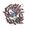
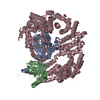
Entry Database : PDB / ID : 4wpnTitle Structure of human ALDH1A1 with inhibitor CM053 Retinal dehydrogenase 1 Keywords / / Function / homology Function Domain/homology Component
/ / / / / / / / / / / / / / / / / / / / / / / / / / / / / / / / / / / / / / / / / / / / / / / / / / / / / / / / / / / / / / Biological species Homo sapiens (human)Method / / Resolution : 1.95 Å Authors Morgan, C.A. / Hurley, T.D. Funding support Organization Grant number Country National Institutes of Health/National Institute on Alcohol Abuse and Alcoholism (NIH/NIAAA) R01AA018123
Journal : J.Med.Chem. / Year : 2015Title : Characterization of Two Distinct Structural Classes of Selective Aldehyde Dehydrogenase 1A1 Inhibitors.Authors : Morgan, C.A. / Hurley, T.D. History Deposition Oct 20, 2014 Deposition site / Processing site Revision 1.0 Feb 11, 2015 Provider / Type Revision 1.1 Mar 11, 2015 Group Revision 1.2 Sep 6, 2017 Group Advisory / Author supporting evidence ... Advisory / Author supporting evidence / Database references / Derived calculations / Other / Source and taxonomy / Structure summary Category citation / entity_src_gen ... citation / entity_src_gen / pdbx_audit_support / pdbx_database_status / pdbx_struct_assembly / pdbx_struct_assembly_prop / pdbx_struct_oper_list / pdbx_validate_close_contact / struct_keywords Item _citation.journal_id_CSD / _entity_src_gen.pdbx_alt_source_flag ... _citation.journal_id_CSD / _entity_src_gen.pdbx_alt_source_flag / _pdbx_audit_support.funding_organization / _pdbx_database_status.pdb_format_compatible / _pdbx_struct_assembly.oligomeric_details / _pdbx_struct_assembly_prop.type / _pdbx_struct_assembly_prop.value / _pdbx_struct_oper_list.symmetry_operation / _struct_keywords.text Revision 1.3 Dec 11, 2019 Group / Category / Item Revision 1.4 Dec 27, 2023 Group / Database references / Refinement descriptionCategory chem_comp_atom / chem_comp_bond ... chem_comp_atom / chem_comp_bond / database_2 / refine_hist Item _database_2.pdbx_DOI / _database_2.pdbx_database_accession ... _database_2.pdbx_DOI / _database_2.pdbx_database_accession / _refine_hist.d_res_low / _refine_hist.number_atoms_solvent / _refine_hist.number_atoms_total / _refine_hist.pdbx_number_atoms_ligand / _refine_hist.pdbx_number_atoms_nucleic_acid / _refine_hist.pdbx_number_atoms_protein
Show all Show less
 Movie
Movie Controller


 Open data
 Basic information
 Components
 Keywords
 Function and homology information
 Homo sapiens (human)
 X-RAY DIFFRACTION /
  Authors
 United States, 1items
  Citation
 Structure visualization
 Downloads & links
 Other downloads
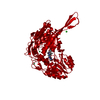


 PDBj
PDBj


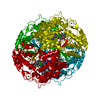
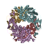
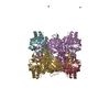
 Assembly
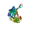


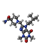
 Mass: 390.480 Da / Num. of mol.: 1 / Source method: obtained synthetically / Formula: C19H30N6O3
 Mass: 35.453 Da / Num. of mol.: 3 / Source method: obtained synthetically / Formula: Cl
 Mass: 173.040 Da / Num. of mol.: 2 / Source method: obtained synthetically / Formula: Yb
 Mass: 18.015 Da / Num. of mol.: 189 / Source method: isolated from a natural source / Formula: H2O
 Sample preparation
 Processing